File:Hertig1956 fig17.jpg: Difference between revisions
mNo edit summary |
m (→Fig. 17.) |
||
| Line 1: | Line 1: | ||
==Fig. 17. | ==Fig. 17. 9-day specimens of Horizon Vb== | ||
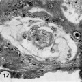
Detail of the embryo, chorionic cavity and adjacent trophoblast. Note lensshaped germ disc, amniogenesis from adjacent eytotrophoblast, and delaminating mesoblasts at the abembryonic pole which are continuous with the endoderm and thus together form the exocoelomic membrane. Carnegie Embryo {{CE8215}}, Section 12-54. X 300. | |||
Three 9-day specimens of Horizon Vb, characterized by syncytiotrophoblastic lacunae with early utero-placental circulation, amniogenesis, a simple bilaminar germ disc and only variable degrees of exocoelomie (Heuser’s) membrane formation. | |||
{{Hertig1956 figures}} | {{Hertig1956 figures}} | ||
[[Category:Carnegie Embryo 8215]] | |||
[[Category:Carnegie Stage 5]] | |||
Latest revision as of 13:36, 31 October 2017
Fig. 17. 9-day specimens of Horizon Vb
Detail of the embryo, chorionic cavity and adjacent trophoblast. Note lensshaped germ disc, amniogenesis from adjacent eytotrophoblast, and delaminating mesoblasts at the abembryonic pole which are continuous with the endoderm and thus together form the exocoelomic membrane. Carnegie Embryo 8215, Section 12-54. X 300.
Three 9-day specimens of Horizon Vb, characterized by syncytiotrophoblastic lacunae with early utero-placental circulation, amniogenesis, a simple bilaminar germ disc and only variable degrees of exocoelomie (Heuser’s) membrane formation.
- Figure Links: 1 | 2 | 3 | 4 | 5 | 6 | 7 | 8 | 9-10 | 11-12 | 13-14 | 15-16 | 17 | 18-19 | 20 | 21-22 | 23 | 24-25 | 26-27 | 28-29 | 30-31 | 32-33 | 34 | 35 | 36 | 37 | 38 | 39 | 40 | 41 | 42 | 43 | 44 | 45 | 46 | 47 | 48 | 40 | 49 | 50 | 51 | 52 | 53 | 54 | 55 | 56 | 57 | 58 | 59 | 60 | 61 | 62 | 63 | 64 | 65 | 66 | 67 | 68 | 69 | 70 | 71 | 72 | 73 | 74 | 75 | 76 | 77 | 78 | 79 | 80 | 81 | 82 | 83 | 84 | 85 | 86 | 87 | 88 | 89 | 90 | plate 1 | plate 2 | plate 3 | plate 4 | plate 5 | plate 6 | plate 7 | plate 8 | plate 9 | plate 10 | plate 11 | plate 12 | plate 13 | plate 14 | plate 15 | plate 16 | plate 17 | table 1 | table 1 image | table 2 image | table 3 image | table 4 | table 4 image | table 5 | table 5 image | All figures | 1956 Hertig | Embryology History - Arthur Hertig | John Rock | Historic Papers
Reference
Hertig AT. Rock J. and Adams EC. A description of 34 human ova within the first 17 days of development. (1956) Amer. J Anat., 98:435-493.
Cite this page: Hill, M.A. (2024, June 27) Embryology Hertig1956 fig17.jpg. Retrieved from https://embryology.med.unsw.edu.au/embryology/index.php/File:Hertig1956_fig17.jpg
- © Dr Mark Hill 2024, UNSW Embryology ISBN: 978 0 7334 2609 4 - UNSW CRICOS Provider Code No. 00098G
File history
Yi efo/eka'e gwa ebo wo le nyangagi wuncin ye kamina wunga tinya nan
| Gwalagizhi | Nyangagi | Dimensions | User | Comment | |
|---|---|---|---|---|---|
| current | 20:22, 23 February 2017 |  | 730 × 726 (100 KB) | Z8600021 (talk | contribs) |
You cannot overwrite this file.
File usage
The following 3 pages use this file: