File:Hertig1956 fig54.jpg: Difference between revisions
mNo edit summary |
mNo edit summary |
||
| Line 1: | Line 1: | ||
==Fig. 54 Reconstruction of half a 7-day ovum== | |||
Which presents an uneven surface of its solid trophohlast (stippled) to the adjacent endometrium. The trophoblast has already surrounded a. capillary sinusoid (left). The cut surface of the hilaminar germ disc is evident (endoderm cellular, ectoderm solid). Note primitive amniogenic cells above tiny amniotic cavity. The maternal sinusoidal network is merely the arteriovenous capillary network of the endometrium that has begun to undergo hypertrophy and dilation. See figures I1 and 12. [[:Category:Carnegie Embryo 8020|Carnegie 8020]]. X 260. | |||
{{Hertig1956 figures}} | {{Hertig1956 figures}} | ||
Latest revision as of 13:24, 24 February 2017
Fig. 54 Reconstruction of half a 7-day ovum
Which presents an uneven surface of its solid trophohlast (stippled) to the adjacent endometrium. The trophoblast has already surrounded a. capillary sinusoid (left). The cut surface of the hilaminar germ disc is evident (endoderm cellular, ectoderm solid). Note primitive amniogenic cells above tiny amniotic cavity. The maternal sinusoidal network is merely the arteriovenous capillary network of the endometrium that has begun to undergo hypertrophy and dilation. See figures I1 and 12. Carnegie 8020. X 260.
- Figure Links: 1 | 2 | 3 | 4 | 5 | 6 | 7 | 8 | 9-10 | 11-12 | 13-14 | 15-16 | 17 | 18-19 | 20 | 21-22 | 23 | 24-25 | 26-27 | 28-29 | 30-31 | 32-33 | 34 | 35 | 36 | 37 | 38 | 39 | 40 | 41 | 42 | 43 | 44 | 45 | 46 | 47 | 48 | 40 | 49 | 50 | 51 | 52 | 53 | 54 | 55 | 56 | 57 | 58 | 59 | 60 | 61 | 62 | 63 | 64 | 65 | 66 | 67 | 68 | 69 | 70 | 71 | 72 | 73 | 74 | 75 | 76 | 77 | 78 | 79 | 80 | 81 | 82 | 83 | 84 | 85 | 86 | 87 | 88 | 89 | 90 | plate 1 | plate 2 | plate 3 | plate 4 | plate 5 | plate 6 | plate 7 | plate 8 | plate 9 | plate 10 | plate 11 | plate 12 | plate 13 | plate 14 | plate 15 | plate 16 | plate 17 | table 1 | table 1 image | table 2 image | table 3 image | table 4 | table 4 image | table 5 | table 5 image | All figures | 1956 Hertig | Embryology History - Arthur Hertig | John Rock | Historic Papers
Reference
Hertig AT. Rock J. and Adams EC. A description of 34 human ova within the first 17 days of development. (1956) Amer. J Anat., 98:435-493.
Cite this page: Hill, M.A. (2024, June 27) Embryology Hertig1956 fig54.jpg. Retrieved from https://embryology.med.unsw.edu.au/embryology/index.php/File:Hertig1956_fig54.jpg
- © Dr Mark Hill 2024, UNSW Embryology ISBN: 978 0 7334 2609 4 - UNSW CRICOS Provider Code No. 00098G
File history
Yi efo/eka'e gwa ebo wo le nyangagi wuncin ye kamina wunga tinya nan
| Gwalagizhi | Nyangagi | Dimensions | User | Comment | |
|---|---|---|---|---|---|
| current | 22:43, 23 February 2017 | 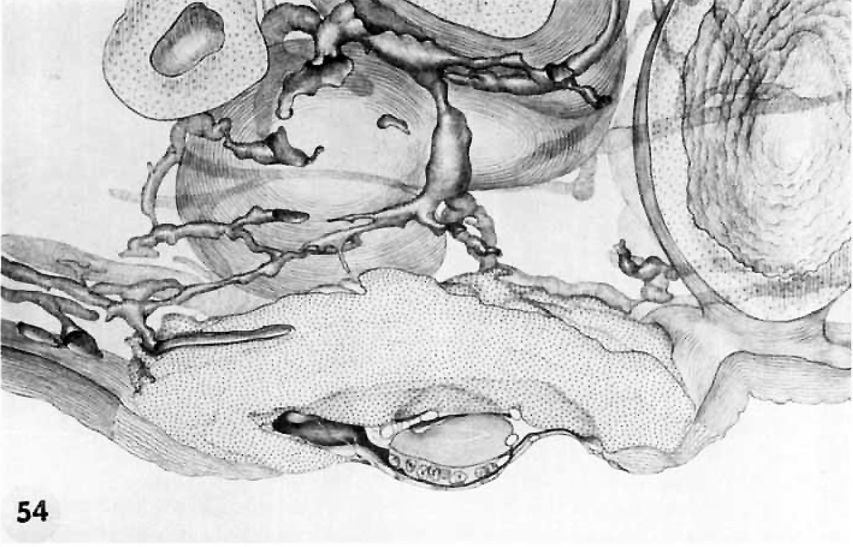 | 1,218 × 782 (185 KB) | Z8600021 (talk | contribs) |
You cannot overwrite this file.
File usage
The following 2 pages use this file: