File:Streeter1915 fig16.jpg: Difference between revisions
mNo edit summary |
mNo edit summary |
||
| Line 12: | Line 12: | ||
{{Footer}} | {{Footer}} | ||
[[Category:Carnegie Embryo | [[Category:Carnegie Embryo 234a]] | ||
Latest revision as of 16:12, 8 February 2017
Figs. 10-17 Simplified profile drawings of the dural veins showing the principal stages in their development in human embryos from 4 mm to birth
It is of particular interest to notice their adaptation to the growth and changes in form of the central nervous system. figure 10, Embryo No. 588, 4 mm.; figure 11, Embryo No. 940, 14 mm.; figure 12, Embryo No. 144, 18 mm.; figure 13, Embryo No. 460, 21 mm.; figure 14, Embryo No. 199, 35 mm.; figure 15, embryo No. 96, 50 mm. crown—rump length; figure 16, Embryo No. 234 a, 80 mm. crown-rump length; figure 17, adult.
| Historic Disclaimer - information about historic embryology pages |
|---|
 |
Links: fig 1 | fig 2 | fig 3 | fig 4 | fig 5 | fig 6 | fig 7 | fig 8 | fig 9 | fig 10 | fig 11 | fig 12 | fig 13 | fig 14 | fig 15 | fig 16 | fig 17 | Streeter 1915 | Neural Vascular | Historic Papers
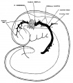
fig 1 embryo 4 mm No. 588
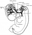
fig 2 embryo 13.8 mm No. 940
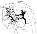
fig 3 embryo 18 mm No. 144
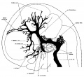
fig 4 embryo 21 mm No. 460
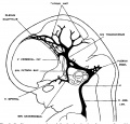
fig 5 embryo 24 mm No. 632
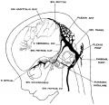
fig 6 embryo 50 mm No. 96
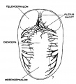
fig 7 embryo 13.8 mm No. 940
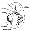
fig 8 embryo 20 mm No. 349
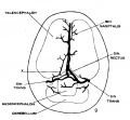
fig 9 fetus 54 mm No. 458
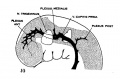
fig 10 embryo 4 mm No. 588
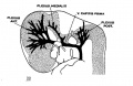
fig 11 embryo 14 mm No. 940
fig 12 embryo 18 mm No. 144
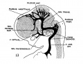
fig 13 embryo 21 mm No. 460
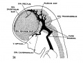
fig 14 embryo 35 mm No. 100
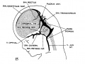
fig 15 embryo 50 mm No. 96
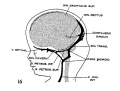
fig 16 embryo 80 mm No. 234a
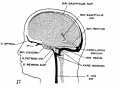
fig 17 adult


Reference
Streeter GL. The development of the venous sinuses of the dura mater in the human embryo. (1915) Amer. J Anat.18: 145-178.
Cite this page: Hill, M.A. (2024, June 26) Embryology Streeter1915 fig16.jpg. Retrieved from https://embryology.med.unsw.edu.au/embryology/index.php/File:Streeter1915_fig16.jpg
- © Dr Mark Hill 2024, UNSW Embryology ISBN: 978 0 7334 2609 4 - UNSW CRICOS Provider Code No. 00098G
File history
Yi efo/eka'e gwa ebo wo le nyangagi wuncin ye kamina wunga tinya nan
| Gwalagizhi | Nyangagi | Dimensions | User | Comment | |
|---|---|---|---|---|---|
| current | 11:20, 26 November 2016 |  | 700 × 533 (76 KB) | Z8600021 (talk | contribs) | {{Historic Disclaimer}} {{Streeter1915 figures}} ===Reference=== {{Ref-Streeter1915}} {{Footer}} Category:Carnegie Embryo 588 |
You cannot overwrite this file.
File usage
The following 21 pages use this file:
- Neural - Meninges Development
- Paper - The development of the venous sinuses of the dura mater in the human embryo
- File:Streeter1915 fig01.jpg
- File:Streeter1915 fig02.jpg
- File:Streeter1915 fig03.jpg
- File:Streeter1915 fig04.jpg
- File:Streeter1915 fig05.jpg
- File:Streeter1915 fig06.jpg
- File:Streeter1915 fig07.jpg
- File:Streeter1915 fig08.jpg
- File:Streeter1915 fig09.jpg
- File:Streeter1915 fig10.jpg
- File:Streeter1915 fig11.jpg
- File:Streeter1915 fig12.jpg
- File:Streeter1915 fig13.jpg
- File:Streeter1915 fig14.jpg
- File:Streeter1915 fig15.jpg
- File:Streeter1915 fig16.jpg
- File:Streeter1915 fig17.jpg
- File:Streeter1915 table01.jpg
- Template:Streeter1915 figures