File:Frazer1910 fig14.jpg: Difference between revisions
(Z8600021 uploaded a new version of File:Frazer1910 fig14.jpg) |
mNo edit summary |
||
| Line 1: | Line 1: | ||
==Fig. 14. Transverse section through this part of the whole larynx== | |||
A transverse section through this part of the whole larynx would now give an outline to the cavity which is shown as a solid diagram in fig. 14; the transverse cavity is bounded in front by the central mass (now becoming more definitely an epiglottis) and is continuous with the sagittal cleft between the two lateral masses, which fit into the angles formed by the junction of the limbs and central stem of the T-shaped cavity, and thus form the two parts of its dorsal wall. | |||
{{Frazer1910 figures}} | {{Frazer1910 figures}} | ||
Latest revision as of 09:46, 11 January 2017
Fig. 14. Transverse section through this part of the whole larynx
A transverse section through this part of the whole larynx would now give an outline to the cavity which is shown as a solid diagram in fig. 14; the transverse cavity is bounded in front by the central mass (now becoming more definitely an epiglottis) and is continuous with the sagittal cleft between the two lateral masses, which fit into the angles formed by the junction of the limbs and central stem of the T-shaped cavity, and thus form the two parts of its dorsal wall.
| Historic Disclaimer - information about historic embryology pages |
|---|
 |
- Links: fig 1 | fig 2 | fig 3 | fig 4 | fig 5 | fig 6 | fig 7 | fig 8 | fig 9 | fig 10 | fig 11 | fig 12 | fig 13 | fig 14 | fig 15 | fig 16 | fig 17 | fig 18 | fig 19 | 1910 Frazer | Historic Embryology Papers | Respiratory System Development
Reference
Frazer JE. Development of the larynx. (1910) J Anat. 44: 156-191. PMID 17232839
Cite this page: Hill, M.A. (2024, June 16) Embryology Frazer1910 fig14.jpg. Retrieved from https://embryology.med.unsw.edu.au/embryology/index.php/File:Frazer1910_fig14.jpg
- © Dr Mark Hill 2024, UNSW Embryology ISBN: 978 0 7334 2609 4 - UNSW CRICOS Provider Code No. 00098G
File history
Click on a date/time to view the file as it appeared at that time.
| Date/Time | Thumbnail | Dimensions | User | Comment | |
|---|---|---|---|---|---|
| current | 09:44, 11 January 2017 | 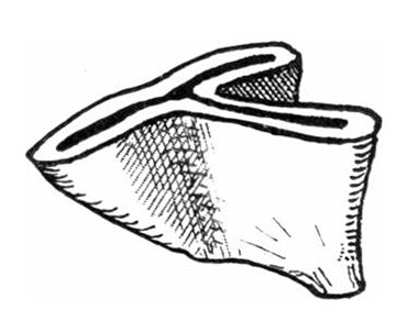 | 380 × 302 (19 KB) | Z8600021 (talk | contribs) | |
| 09:44, 11 January 2017 |  | 417 × 385 (19 KB) | Z8600021 (talk | contribs) | {{Frazer1910 figures}} |
You cannot overwrite this file.
File usage
The following 2 pages use this file: