File:Jenkinson1913 fig149.jpg: Difference between revisions
(Z8600021 uploaded a new version of File:Jenkinson1913 fig149.jpg) |
mNo edit summary |
||
| Line 1: | Line 1: | ||
==Fig. 149. Five stages in the formation of the placenta in the mouse== | |||
A, The blastocyst free in the uterus. | |||
B, The blastocyst attached and the placental thickening of the a1lantoidean trophoblast developed (a.tr.). | |||
C, Later stage, after elosure of the amniotic cavity (am.c.). | |||
D, The foetal blood-vessels beginning to penetrate the allantoidean trophoblast. | |||
E, Elaboration of the placenta. Disappearance of the distal Wall of the yolk-sac and omphaloidean trophoblast (o.tr.). | |||
l’.u’., new uterine lumen on the antimesometrie side; l.u., original lumen of the uterus ; 3/.s., yolk-sac ; g/.st. yolkstalk ; u.c., umbilical cord; m., mesometrium. | |||
{{Historic Disclaimer}} | {{Historic Disclaimer}} | ||
===Reference=== | ===Reference=== | ||
| Line 4: | Line 19: | ||
{{Footer}} | {{Footer}} | ||
[[Category:Mouse]] | |||
Latest revision as of 14:06, 15 December 2016
Fig. 149. Five stages in the formation of the placenta in the mouse
A, The blastocyst free in the uterus.
B, The blastocyst attached and the placental thickening of the a1lantoidean trophoblast developed (a.tr.).
C, Later stage, after elosure of the amniotic cavity (am.c.).
D, The foetal blood-vessels beginning to penetrate the allantoidean trophoblast.
E, Elaboration of the placenta. Disappearance of the distal Wall of the yolk-sac and omphaloidean trophoblast (o.tr.).
l’.u’., new uterine lumen on the antimesometrie side; l.u., original lumen of the uterus ; 3/.s., yolk-sac ; g/.st. yolkstalk ; u.c., umbilical cord; m., mesometrium.
| Historic Disclaimer - information about historic embryology pages |
|---|
 |
Reference
Jenkinson JW. Vertebrate Embryology. (1913) Oxford University Press, London.
Cite this page: Hill, M.A. (2024, June 18) Embryology Jenkinson1913 fig149.jpg. Retrieved from https://embryology.med.unsw.edu.au/embryology/index.php/File:Jenkinson1913_fig149.jpg
- © Dr Mark Hill 2024, UNSW Embryology ISBN: 978 0 7334 2609 4 - UNSW CRICOS Provider Code No. 00098G
File history
Yi efo/eka'e gwa ebo wo le nyangagi wuncin ye kamina wunga tinya nan
| Gwalagizhi | Nyangagi | Dimensions | User | Comment | |
|---|---|---|---|---|---|
| current | 14:00, 15 December 2016 | 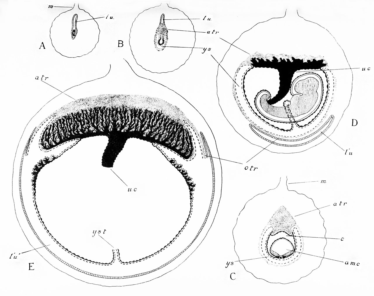 | 1,280 × 1,014 (188 KB) | Z8600021 (talk | contribs) | |
| 13:59, 15 December 2016 |  | 3,025 × 1,897 (622 KB) | Z8600021 (talk | contribs) | {{Historic Disclaimer}} ===Reference=== {{Ref-Jenkinson1913}} {{Footer}} |
You cannot overwrite this file.
File usage
The following 2 pages use this file: