File:Cooper1938 plate30.jpg: Difference between revisions
(==Plate XXXI== Fig. 17. Swollen cells lining an alveolus in the bronchopneumonic lung of an infant aged 12 weeks. X 630. Fig. 18. Cells lining alveoli in the lung of an infant aged 8 month...) |
mNo edit summary |
||
| Line 1: | Line 1: | ||
==Plate XXXI== | ==Plate XXXI== | ||
===Plate XXX=== | |||
[[:File:Cooper1938 | [[:File:Cooper1938 fig14.jpg|Fig. 14]]. Surface view of alveolar cells. Cap. = capillary; R.B.C. : erythrocyte. Full term embryo. X IVU. | ||
[[:File:Cooper1938 | [[:File:Cooper1938 fig15.jpg|Fig. 15]]. Flattened and cubical cells lining alveoli. A : flattened cells; B : cubical cells. Full term embryo. X 700. | ||
[[:File:Cooper1938 fig16.jpg|Fig. 16]]. Cells lining the alveoli in the lung of an anencephalic full - term foetus which had breathed. Note continuity of cellular lining of alveolus marked X. X300. | |||
<gallery> | <gallery> | ||
File:Cooper1938 | File:Cooper1938 fig14.jpg|Fig. 17. alveolar cells Full term embryo | ||
File:Cooper1938 | File:Cooper1938 fig15.jpg|Fig. 18. Flattened and cubical cells Full term embryo | ||
File:Cooper1938 | File:Cooper1938 fig16.jpg|Fig. 16. alveoli term foetus | ||
</gallery> | </gallery> | ||
Revision as of 10:16, 27 November 2016
Plate XXXI
Plate XXX
Fig. 14. Surface view of alveolar cells. Cap. = capillary; R.B.C. : erythrocyte. Full term embryo. X IVU.
Fig. 15. Flattened and cubical cells lining alveoli. A : flattened cells; B : cubical cells. Full term embryo. X 700.
Fig. 16. Cells lining the alveoli in the lung of an anencephalic full - term foetus which had breathed. Note continuity of cellular lining of alveolus marked X. X300.
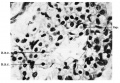
Fig. 17. alveolar cells Full term embryo
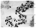
Fig. 18. Flattened and cubical cells Full term embryo
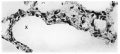
Fig. 16. alveoli term foetus


| Historic Disclaimer - information about historic embryology pages |
|---|
 |
Reference
Cooper ERA. A histological investigation of the development and structure of the human lung. (1938) J Pathology 47: 105-114.
Cite this page: Hill, M.A. (2024, June 21) Embryology Cooper1938 plate30.jpg. Retrieved from https://embryology.med.unsw.edu.au/embryology/index.php/File:Cooper1938_plate30.jpg
- © Dr Mark Hill 2024, UNSW Embryology ISBN: 978 0 7334 2609 4 - UNSW CRICOS Provider Code No. 00098G
File history
Yi efo/eka'e gwa ebo wo le nyangagi wuncin ye kamina wunga tinya nan
| Gwalagizhi | Nyangagi | Dimensions | User | Comment | |
|---|---|---|---|---|---|
| current | 10:12, 27 November 2016 |  | 1,655 × 2,457 (402 KB) | Z8600021 (talk | contribs) | ==Plate XXXI== Fig. 17. Swollen cells lining an alveolus in the bronchopneumonic lung of an infant aged 12 weeks. X 630. Fig. 18. Cells lining alveoli in the lung of an infant aged 8 month... |
You cannot overwrite this file.
File usage
The following page uses this file: