File:Stages Plasmodium falciparum.jpg: Difference between revisions
From Embryology
(Stages of Plasmodium falciparum as drawn from microscopic observation of thin blood smears (1971). Fig. 1: Normal red cell Figs. 2-18: Trophozoites (among these, Figs. 2-10 correspond to ring-stag trophozoites) Figs. 19-26: Schizonts (Fig. 26 is a rupt) |
No edit summary |
||
| Line 13: | Line 13: | ||
Illustrations from: Coatney GR, Collins WE, Warren M, Contacos PG. The Primate Malarias. Bethesda: U.S. Department of Health, Education and Welfare; 1971. | Illustrations from: Coatney GR, Collins WE, Warren M, Contacos PG. The Primate Malarias. Bethesda: U.S. Department of Health, Education and Welfare; 1971. | ||
From CDC: [http://www.dpd.cdc.gov/dpdx/hTML/Frames/M-R/Malaria/falciparum/body_malariadffalcipar.htm Diagnostic findings - Historic Images] | |||
[[Category:Cartoon]] | [[Category:Cartoon]] | ||
Latest revision as of 01:09, 26 May 2010
Stages of Plasmodium falciparum as drawn from microscopic observation of thin blood smears (1971).
Fig. 1: Normal red cell
Figs. 2-18: Trophozoites (among these, Figs. 2-10 correspond to ring-stag trophozoites)
Figs. 19-26: Schizonts (Fig. 26 is a ruptured schizont)
Figs.27, 28: Mature macrogametocytes (female)
Figs. 29, 30: Mature microgametocytes (male)
Illustrations from: Coatney GR, Collins WE, Warren M, Contacos PG. The Primate Malarias. Bethesda: U.S. Department of Health, Education and Welfare; 1971.
From CDC: Diagnostic findings - Historic Images
File history
Yi efo/eka'e gwa ebo wo le nyangagi wuncin ye kamina wunga tinya nan
| Gwalagizhi | Nyangagi | Dimensions | User | Comment | |
|---|---|---|---|---|---|
| current | 01:09, 26 May 2010 | 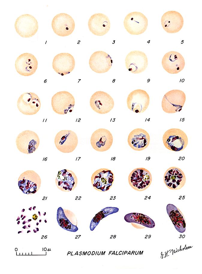 | 674 × 914 (65 KB) | S8600021 (talk | contribs) | Stages of Plasmodium falciparum as drawn from microscopic observation of thin blood smears (1971). Fig. 1: Normal red cell Figs. 2-18: Trophozoites (among these, Figs. 2-10 correspond to ring-stag trophozoites) Figs. 19-26: Schizonts (Fig. 26 is a rupt |
You cannot overwrite this file.
File usage
There are no pages that use this file.