File:Complete hydatidiform mole 05.jpg: Difference between revisions
From Embryology
mNo edit summary |
mNo edit summary |
||
| Line 3: | Line 3: | ||
===Reference=== | ===Reference=== | ||
{{SOC}} | {{SOC embryo}} | ||
{{Footer}} | {{Footer}} | ||
[[Category:Human]] [[Category:Abnormal Development]] [[Category:Uterus]][[Category:Hydatidiform Mole]] | [[Category:Human]] [[Category:Abnormal Development]] [[Category:Uterus]][[Category:Hydatidiform Mole]] | ||
Revision as of 22:47, 31 May 2016
- Links: Hydatidiform Mole
Reference
Image: Dr Steven O'Connor (Houston, Texas) - Other embryo images.
Cite this page: Hill, M.A. (2024, June 26) Embryology Complete hydatidiform mole 05.jpg. Retrieved from https://embryology.med.unsw.edu.au/embryology/index.php/File:Complete_hydatidiform_mole_05.jpg
- © Dr Mark Hill 2024, UNSW Embryology ISBN: 978 0 7334 2609 4 - UNSW CRICOS Provider Code No. 00098G
File history
Yi efo/eka'e gwa ebo wo le nyangagi wuncin ye kamina wunga tinya nan
| Gwalagizhi | Nyangagi | Dimensions | User | Comment | |
|---|---|---|---|---|---|
| current | 22:45, 31 May 2016 | 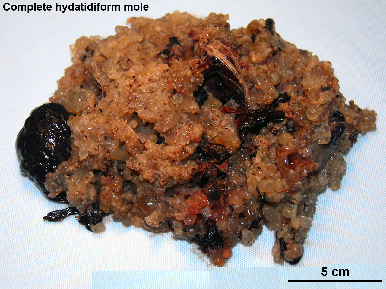 | 1,280 × 960 (324 KB) | Z8600021 (talk | contribs) |
You cannot overwrite this file.
File usage
The following page uses this file: