File:Human week 10 fetus 09.jpg: Difference between revisions
mNo edit summary |
|||
| Line 5: | Line 5: | ||
Note the atlas cervical vertebra (C1) and axis cervical vertebra (C2) specialised vertebra of the axial skeleton to connect the skull to the vertebral column. | Note the atlas cervical vertebra (C1) and axis cervical vertebra (C2) specialised vertebra of the axial skeleton to connect the skull to the vertebral column. | ||
The axis odontoid process is shown. Note that at this stage the vertebral column is still cartilage. | |||
{{Human Female Fetus Week 10 gallery}} | {{Human Female Fetus Week 10 gallery}} | ||
Revision as of 14:44, 25 May 2016
Human Female Fetus Atlas and Axis (10 week)
Large image version of plane D, close to midline (Stain - Haematoxylin Eosin). 0.5 mm scale bar
Note the atlas cervical vertebra (C1) and axis cervical vertebra (C2) specialised vertebra of the axial skeleton to connect the skull to the vertebral column.
The axis odontoid process is shown. Note that at this stage the vertebral column is still cartilage.
- Human Female Fetus (week 10)
Sagittal Section (plane D)

Pituitary and Lamina Terminalis

Olfactory Nerve

Atlas and Axis

Sacrum

Oral Cavity

Epiglottis

Heart

Spleen

Midgut Herniation

Midgut Herniation (label)

Pelvic Region

Pelvic Region (label)


Related Images
Fetus (week 10) Planes A (most lateral), B (lateral), C (medial) and D (midline) from lateral towards the midline.
- Human Fetus - most lateral | lateral | medial | midline
- Head - most lateral | lateral | medial | midline
- Cerebellum - most lateral | lateral | medial | midline
- Urogenital Unlabelled - most lateral | lateral | medial | midline
- Urogenital Labelled - most lateral | lateral | medial | midline
- Large Images - midline
- Image Source: UNSW Embryology, no reproduction without permission.
File history
Yi efo/eka'e gwa ebo wo le nyangagi wuncin ye kamina wunga tinya nan
| Gwalagizhi | Nyangagi | Dimensions | User | Comment | |
|---|---|---|---|---|---|
| current | 23:00, 17 June 2012 | 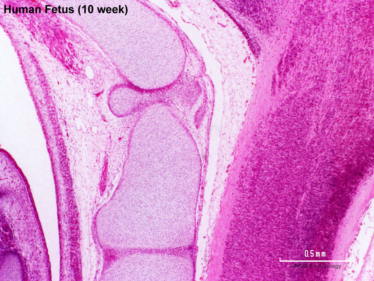 | 1,200 × 900 (345 KB) | Z8600021 (talk | contribs) | ==Human Female Fetus Atlas and Axis (10 week)== Large image version of plane D, close to midline (H&E stain). 0.5 mm scale bar Note: {{10wkFetus}} |
You cannot overwrite this file.
File usage
The following 17 pages use this file:
- BGDA Practical 12 - Embryo to Fetus
- Fetal Development - 10 Weeks
- Foundations Practical - Week 9 to 36
- File:Human week 10 fetus 01.jpg
- File:Human week 10 fetus 03.jpg
- File:Human week 10 fetus 04.jpg
- File:Human week 10 fetus 05.jpg
- File:Human week 10 fetus 06.jpg
- File:Human week 10 fetus 07.jpg
- File:Human week 10 fetus 08.jpg
- File:Human week 10 fetus 09.jpg
- File:Human week 10 fetus 10.jpg
- File:Human week 10 fetus 11.jpg
- File:Human week 10 fetus 12.jpg
- File:Human week 10 fetus 23.jpg
- File:Human week 10 fetus 26.jpg
- Template:Human Female Fetus Week 10 gallery