File:Bardeen1906-plate32.jpg: Difference between revisions
From Embryology
No edit summary |
mNo edit summary |
||
| Line 1: | Line 1: | ||
==Plate III. Human Embryo 11 mm== | |||
Three figures to illustrate the skeletal, muscular, and nervous apparatus of the right posterior extremity of embryo CIX, length 11 mm., age about five Weeks. The nerves are represented black; the muscle anlages by stippling; the skeletal structures, light grey; the skin of the leg, transparent. About 17 diam. | |||
FIG. 2. Lateral view. | |||
FIG. 3. Ventral view showing the relation of the pelvis to the body wall and the main nerve trunks. At the right the outline of the peritoneal membrane is shown. The division of the main nerve trunks into branches is diagrammatic. | |||
Revision as of 21:50, 7 September 2015
Plate III. Human Embryo 11 mm
Three figures to illustrate the skeletal, muscular, and nervous apparatus of the right posterior extremity of embryo CIX, length 11 mm., age about five Weeks. The nerves are represented black; the muscle anlages by stippling; the skeletal structures, light grey; the skin of the leg, transparent. About 17 diam.
FIG. 2. Lateral view.
FIG. 3. Ventral view showing the relation of the pelvis to the body wall and the main nerve trunks. At the right the outline of the peritoneal membrane is shown. The division of the main nerve trunks into branches is diagrammatic.
File history
Yi efo/eka'e gwa ebo wo le nyangagi wuncin ye kamina wunga tinya nan
| Gwalagizhi | Nyangagi | Dimensions | User | Comment | |
|---|---|---|---|---|---|
| current | 21:47, 7 September 2015 | 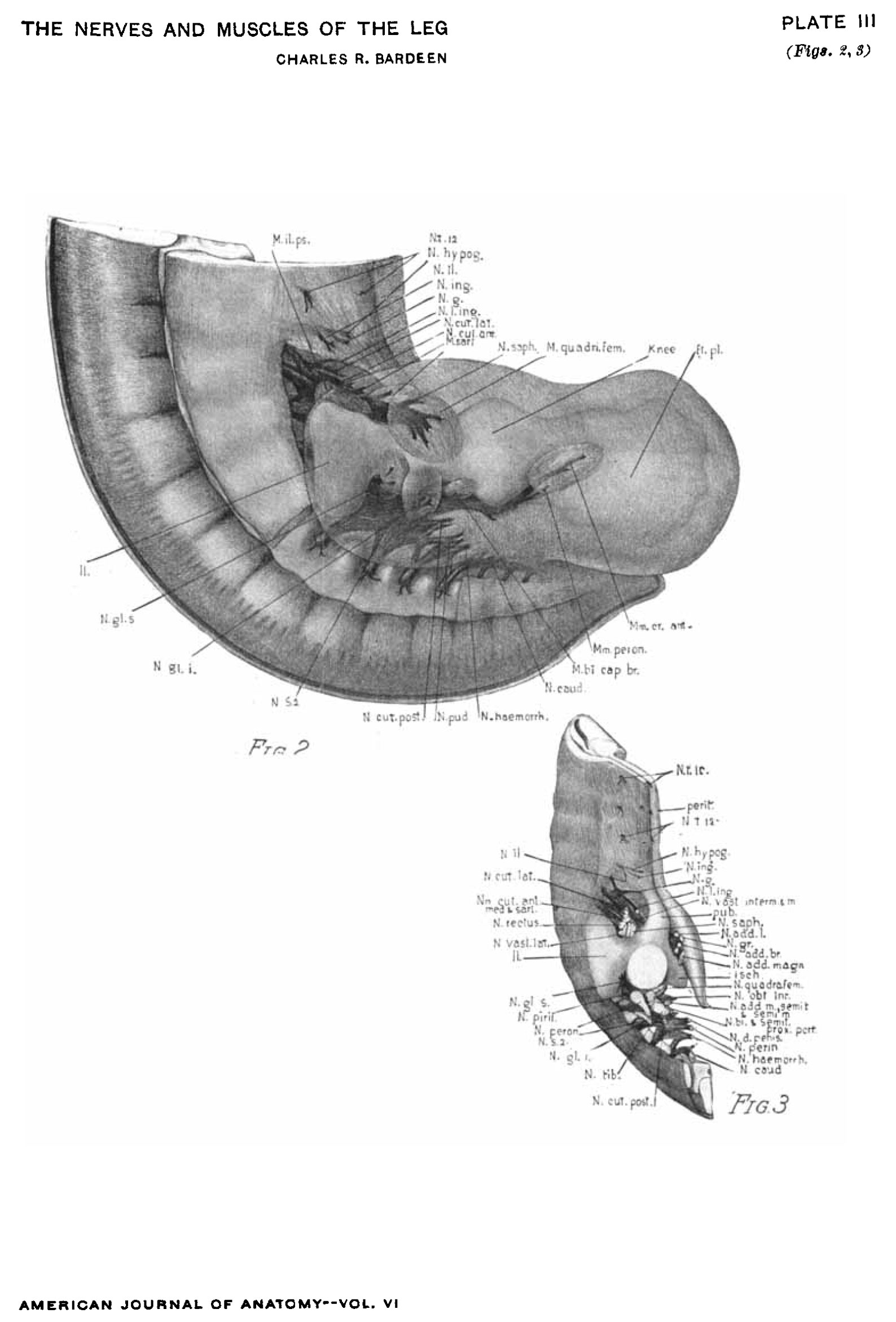 | 1,588 × 2,341 (292 KB) | Z8600021 (talk | contribs) |
You cannot overwrite this file.
File usage
The following page uses this file: