File:DibleWest1941 fig03.jpg: Difference between revisions
mNo edit summary |
mNo edit summary |
||
| Line 1: | Line 1: | ||
==Fig. 3. Coloured drawing of a section (slide 25-2) through the middle of the ovum== | ==Fig. 3. Coloured drawing of a section (slide 25-2) through the middle of the ovum== | ||
The ovum (Fig. 3) consists of a slightly flattened chorionic vesicle, the blastocyst, which has an internal measurement of 047mm. in the equatorial plane, i.e. from side to side, and of 0.28 mm. in the polar plane, i.e. from the surface inward. The inner surface of the blastocyst is smooth and has the embryonic rudiment attached at its base; the outer surface is irregular owing to cellular and syncytial processes growing out from it into the implantation cavity. | |||
Latest revision as of 23:16, 31 July 2015
Fig. 3. Coloured drawing of a section (slide 25-2) through the middle of the ovum
The ovum (Fig. 3) consists of a slightly flattened chorionic vesicle, the blastocyst, which has an internal measurement of 047mm. in the equatorial plane, i.e. from side to side, and of 0.28 mm. in the polar plane, i.e. from the surface inward. The inner surface of the blastocyst is smooth and has the embryonic rudiment attached at its base; the outer surface is irregular owing to cellular and syncytial processes growing out from it into the implantation cavity.
x100.
| Historic Disclaimer - information about historic embryology pages |
|---|
 |
- Links: Fig. 1 | Fig. 2 | Fig. 3 | Fig. 4 | Fig. 5 | Fig. 6 | Fig. 7 | Fig. 8 | Fig. 9 | Fig. 10 | Fig. 11 | Plate 1 | Plate 2 | Plate 3 | Plate 4 | Plate 5 | Plate 6
Reference
Dible JH. and West CM. A human ovum at the previllous stage. (1941) J Anat. 75(3): 269–281. PMID 17104860
Cite this page: Hill, M.A. (2024, June 27) Embryology DibleWest1941 fig03.jpg. Retrieved from https://embryology.med.unsw.edu.au/embryology/index.php/File:DibleWest1941_fig03.jpg
- © Dr Mark Hill 2024, UNSW Embryology ISBN: 978 0 7334 2609 4 - UNSW CRICOS Provider Code No. 00098G
File history
Yi efo/eka'e gwa ebo wo le nyangagi wuncin ye kamina wunga tinya nan
| Gwalagizhi | Nyangagi | Dimensions | User | Comment | |
|---|---|---|---|---|---|
| current | 16:33, 28 July 2015 | 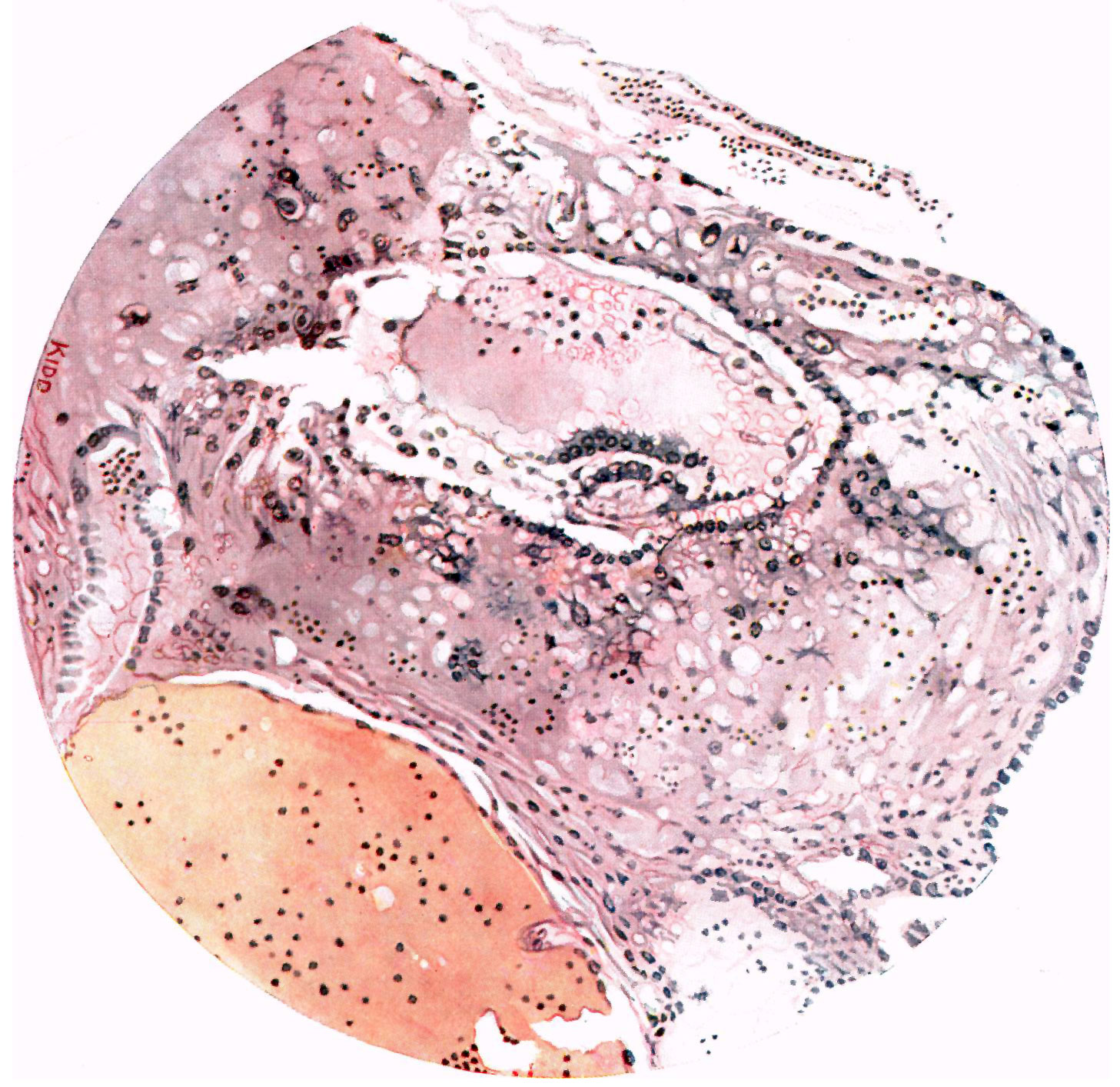 | 1,457 × 1,413 (458 KB) | Z8600021 (talk | contribs) |
You cannot overwrite this file.
File usage
The following page uses this file: