File:Gray0461.jpg: Difference between revisions
(Z8600021 uploaded a new version of "File:Gray0461.jpg") |
mNo edit summary |
||
| Line 1: | Line 1: | ||
==Fig. 461. Diagram to illustrate the simple tubular condition of the heart== | |||
(Drawn from Ecker-Ziegler model.) | |||
The intermediate portion arches transversely from left to right, and then turns sharply forward into the anterior part of the loop. Slight constrictions make their appearance in the tube and divide it from behind forward into five parts, viz.: (1) the sinus venosus; (2) the primitive atrium; (3) the primitive ventricle; (4) the bulbus cordis, and (5) the truncus arteriosus (Figs. 461, 462). The constriction between the atrium and ventricle constitutes the atrial canal, and indicates the site of the future atrioventricular valves. | |||
{{Historic Disclaimer}} | |||
{{Gray Anatomy}} | |||
[[Category:Cardiovascular]] [[Category:Blood Vessel]][Category:Chicken]] | |||
[[Category:Historic Embryology]] | |||
[[Category:Gray's 1918 Anatomy]] | |||
Latest revision as of 18:34, 23 August 2014
Fig. 461. Diagram to illustrate the simple tubular condition of the heart
(Drawn from Ecker-Ziegler model.)
The intermediate portion arches transversely from left to right, and then turns sharply forward into the anterior part of the loop. Slight constrictions make their appearance in the tube and divide it from behind forward into five parts, viz.: (1) the sinus venosus; (2) the primitive atrium; (3) the primitive ventricle; (4) the bulbus cordis, and (5) the truncus arteriosus (Figs. 461, 462). The constriction between the atrium and ventricle constitutes the atrial canal, and indicates the site of the future atrioventricular valves.
| Historic Disclaimer - information about historic embryology pages |
|---|
 |
- Gray's Images: Development | Lymphatic | Neural | Vision | Hearing | Somatosensory | Integumentary | Respiratory | Gastrointestinal | Urogenital | Endocrine | Surface Anatomy | iBook | Historic Disclaimer
| Historic Disclaimer - information about historic embryology pages |
|---|
|
| iBook - Gray's Embryology | |
|---|---|

|
|
Reference
Gray H. Anatomy of the human body. (1918) Philadelphia: Lea & Febiger.
Cite this page: Hill, M.A. (2024, June 26) Embryology Gray0461.jpg. Retrieved from https://embryology.med.unsw.edu.au/embryology/index.php/File:Gray0461.jpg
- © Dr Mark Hill 2024, UNSW Embryology ISBN: 978 0 7334 2609 4 - UNSW CRICOS Provider Code No. 00098G[Category:Chicken]]
File history
Yi efo/eka'e gwa ebo wo le nyangagi wuncin ye kamina wunga tinya nan
| Gwalagizhi | Nyangagi | Dimensions | User | Comment | |
|---|---|---|---|---|---|
| current | 18:29, 23 August 2014 | 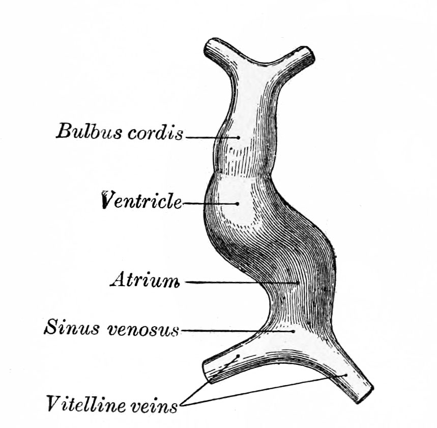 | 859 × 841 (82 KB) | Z8600021 (talk | contribs) | |
| 18:29, 23 August 2014 | 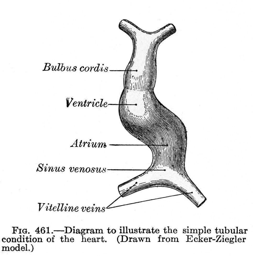 | 966 × 980 (110 KB) | Z8600021 (talk | contribs) |
You cannot overwrite this file.
File usage
The following page uses this file: