File:Mouse embryo E11 tomography 01.jpg: Difference between revisions
From Embryology
mNo edit summary |
mNo edit summary |
||
| Line 4: | Line 4: | ||
:'''Links:''' [[Mouse Development]] | [[:Category:Mouse E11.0|Category:Mouse E11.0]] | [[Computed Tomography]] | [[:File:Mouse embryo E11 HNF3beta notochord marker 01.jpg|Image - Mouse embryo E11 | :'''Links:''' [[Mouse Development]] | [[:Category:Mouse E11.0|Category:Mouse E11.0]] | [[Computed Tomography]] | [[:File:Mouse_embryo_E11_and_tomography_01.jpg|Image - Mouse embryo E11 and tomography]] | [[:File:Mouse embryo E11 HNF3beta notochord marker 01.jpg|Image - Mouse embryo E11 notochord marker]] | ||
===Reference=== | ===Reference=== | ||
Revision as of 13:12, 18 August 2014
Mouse Embryo (E11) Optical Projection Tomography
External view of an E11 Theiler stage (TS 18) control embryo righthand side surface rendered view of the Optical Projection Tomography (OPT) 3D reconstruction.
- Links: Mouse Development | Category:Mouse E11.0 | Computed Tomography | Image - Mouse embryo E11 and tomography | Image - Mouse embryo E11 notochord marker
Reference
<pubmed>22132119</pubmed>| PLoS One.
Copyright
© 2011 Hajduk et al. This is an open-access article distributed under the terms of the Creative Commons Attribution License, which permits unrestricted use, distribution, and reproduction in any medium, provided the original author and source are credited.
Figure 1. doi:10.1371/journal.pone.0027635.g001 Panel B cropped from full figure, resized and relabelled.
File history
Yi efo/eka'e gwa ebo wo le nyangagi wuncin ye kamina wunga tinya nan
| Gwalagizhi | Nyangagi | Dimensions | User | Comment | |
|---|---|---|---|---|---|
| current | 13:04, 18 August 2014 | 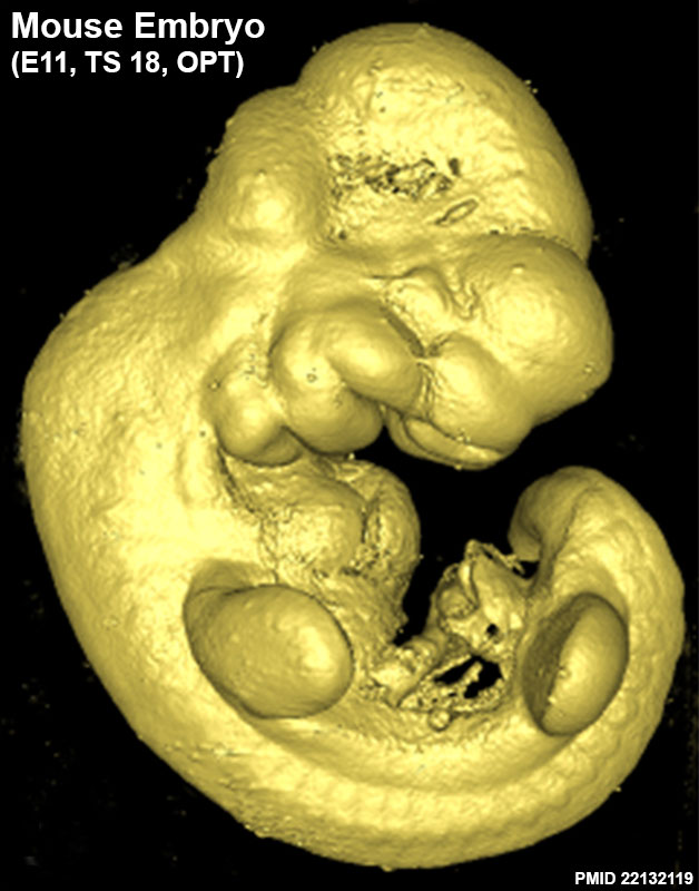 | 628 × 800 (82 KB) | Z8600021 (talk | contribs) |
You cannot overwrite this file.
File usage
There are no pages that use this file.