File:Intestine villi crypts cartoon.jpg: Difference between revisions
From Embryology
(==Intestine Histology== {{Intestine Histology}} {{Blue Histology}}) |
mNo edit summary |
||
| Line 1: | Line 1: | ||
==Intestine Histology== | ==Intestine Histology Cartoon== | ||
===Intestinal Villi=== | |||
* cover entire intestinal mucosa | |||
* about one mm long | |||
* increase the surface area by a factor of ~ ten. | |||
* villi surface formed by a simple columnar epithelium. | |||
* each absorptive cell or '''enterocyte''' of the epithelium forms numerous microvilli | |||
===Intestinal Crypts=== | |||
(crypts of Lieberkühn) | |||
* openings of simple tubular glands | |||
* extend through the lamina propria down to the muscularis mucosae | |||
* undifferentiated cells close to the bottom of the crypts regenerate/replace the epithelium | |||
** epithelial cell turnover time is less than one week | |||
* other epithelial cells in the crypts correspond largely to those in the epithelium of the intestinal villi | |||
* '''Paneth cells''' are located at the bottom of the crypts | |||
** release a number of antibacterial substances (including lysozyme) | |||
** thought to be involved in the control of infections | |||
{{Intestine Histology}} | {{Intestine Histology}} | ||
{{Blue Histology}} | {{Blue Histology}} | ||
Revision as of 13:02, 5 April 2013
Intestine Histology Cartoon
Intestinal Villi
- cover entire intestinal mucosa
- about one mm long
- increase the surface area by a factor of ~ ten.
- villi surface formed by a simple columnar epithelium.
- each absorptive cell or enterocyte of the epithelium forms numerous microvilli
Intestinal Crypts
(crypts of Lieberkühn)
- openings of simple tubular glands
- extend through the lamina propria down to the muscularis mucosae
- undifferentiated cells close to the bottom of the crypts regenerate/replace the epithelium
- epithelial cell turnover time is less than one week
- other epithelial cells in the crypts correspond largely to those in the epithelium of the intestinal villi
- Paneth cells are located at the bottom of the crypts
- release a number of antibacterial substances (including lysozyme)
- thought to be involved in the control of infections
- Intestine Histology Links: Duodenum overview | Duodenum villi and crypts | Duodenum | Jejunum overview | Jejunum villus | Jejunum labeled | Jejunum unlabeled | Gastrointestinal Tract Histology | Intestine Development
Links: Histology | Histology Stains | Blue Histology images copyright Lutz Slomianka 1998-2009. The literary and artistic works on the original Blue Histology website may be reproduced, adapted, published and distributed for non-commercial purposes. See also the page Histology Stains.
Cite this page: Hill, M.A. (2024, June 3) Embryology Intestine villi crypts cartoon.jpg. Retrieved from https://embryology.med.unsw.edu.au/embryology/index.php/File:Intestine_villi_crypts_cartoon.jpg
- © Dr Mark Hill 2024, UNSW Embryology ISBN: 978 0 7334 2609 4 - UNSW CRICOS Provider Code No. 00098G
File history
Click on a date/time to view the file as it appeared at that time.
| Date/Time | Thumbnail | Dimensions | User | Comment | |
|---|---|---|---|---|---|
| current | 12:55, 5 April 2013 | 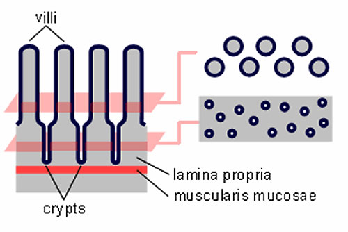 | 500 × 334 (29 KB) | Z8600021 (talk | contribs) | ==Intestine Histology== {{Intestine Histology}} {{Blue Histology}} |
You cannot overwrite this file.