File:Lineback1920 fig04-5.jpg: Difference between revisions
From Embryology
No edit summary |
No edit summary |
||
| Line 1: | Line 1: | ||
Fig 4 Cross-section of the colon of a human fetus 52 mm. CR. length. | |||
Showing a well-defined tenia at the mesenteric arc with a complete layer of loosely scattered fibers surrounding the tube. | |||
Fig 5 Cross-section of a human fetus 90 mm. CR. length. | |||
Showing the fibers of the longitudinal muscle, loosely scattered in the 52-mm. stage, now compact, and only the mesenteric taenia present. Two enlargements are seen, one on each side of the tube, caused by the presence of blood-vessels. It will be noted that the muscle here is in no way involved. | |||
Revision as of 08:50, 27 December 2012
Fig 4 Cross-section of the colon of a human fetus 52 mm. CR. length.
Showing a well-defined tenia at the mesenteric arc with a complete layer of loosely scattered fibers surrounding the tube.
Fig 5 Cross-section of a human fetus 90 mm. CR. length.
Showing the fibers of the longitudinal muscle, loosely scattered in the 52-mm. stage, now compact, and only the mesenteric taenia present. Two enlargements are seen, one on each side of the tube, caused by the presence of blood-vessels. It will be noted that the muscle here is in no way involved.
File history
Yi efo/eka'e gwa ebo wo le nyangagi wuncin ye kamina wunga tinya nan
| Gwalagizhi | Nyangagi | Dimensions | User | Comment | |
|---|---|---|---|---|---|
| current | 08:48, 27 December 2012 | 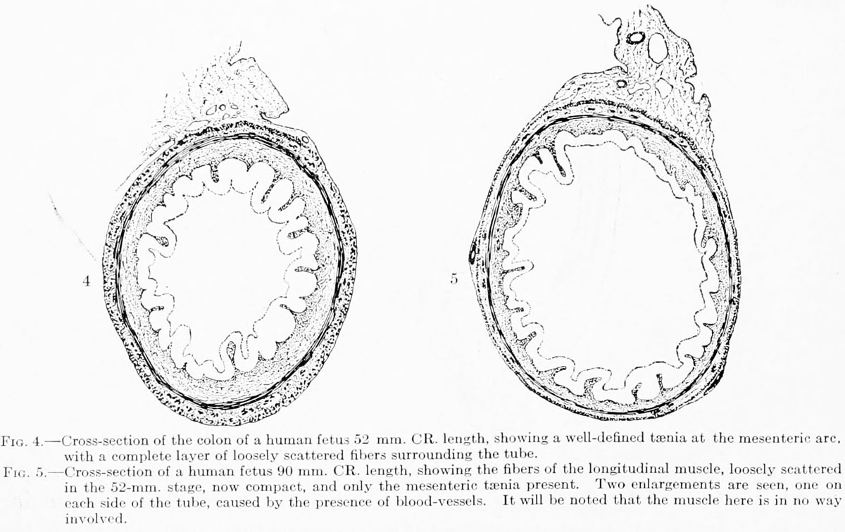 | 1,200 × 756 (150 KB) | Z8600021 (talk | contribs) |
You cannot overwrite this file.
File usage
The following page uses this file: