File:Ossification endochondral 01.jpg: Difference between revisions
From Embryology
No edit summary |
No edit summary |
||
| Line 3: | Line 3: | ||
* Histological image of a developing vertebra (neonatal rat). | * Histological image of a developing vertebra (neonatal rat). | ||
* vertebra - cartilage template and developing bony collar (centre of image) | * vertebra - cartilage template and developing bony collar (centre of image) | ||
===Legend=== | |||
* blue - cartilage matrix | |||
* red - bone matrix | |||
scale bar 160 microns | scale bar 160 microns | ||
Revision as of 12:21, 26 March 2012
Developing Vertebra - Endochondral Ossification
- Histological image of a developing vertebra (neonatal rat).
- vertebra - cartilage template and developing bony collar (centre of image)
Legend
- blue - cartilage matrix
- red - bone matrix
scale bar 160 microns
Original File Name: Endochondral9x5-1000px.jpg
Image Source: UNSW Embryology
File history
Yi efo/eka'e gwa ebo wo le nyangagi wuncin ye kamina wunga tinya nan
| Gwalagizhi | Nyangagi | Dimensions | User | Comment | |
|---|---|---|---|---|---|
| current | 12:33, 23 March 2012 | 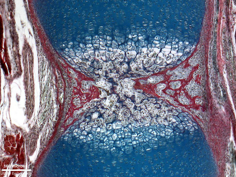 | 817 × 613 (198 KB) | Z8600021 (talk | contribs) | ==Developing Vertebra - Endochondral Ossification== * Histological image of a developing vertebra and intervertebral disc (neonatal rat). * intervertebral disc - nucleus pulposus and annular fibrocartilage (bottom of image) * vertebra - cartilage templa |
You cannot overwrite this file.
File usage
The following 2 pages use this file: