File:Lymph node structure 02.jpg: Difference between revisions
From Embryology
No edit summary |
No edit summary |
||
| Line 1: | Line 1: | ||
==Lymph | ==Lymph Node Structure== | ||
Schematic representation of the organization of a lymph node | Schematic representation of the organization of a lymph node. | ||
* Afferent lymphatics enter lymph nodes and deliver lymph to the subcapsular sinus (SCS), which forms a channel around the periphery of the lymph node. | |||
* Lymphatic sinuses run from the SCS through the cortex to the medulla, and exit the lymph node via efferent lymphatic vessels on the opposite, hilar, side of the organ. * B cell follicles containing follicular dendritic cell (FDC) networks are arranged in the lymph node cortex and are separated from the SCS by a layer of marginal reticular cells (MRC). | |||
* The T cells zones in the paracortex, which contain many fibroblastic reticular cells (FRC), are separated by the cortical ridge, an area rich in T cells, dendritic cells (DCs), blood vessels, and FRC. | |||
* Blood vessels enter and exit the lymph node on the hilar side, and snake through the lymph node like the branches of a tree. | |||
* Specialized high endothelial venules (HEVs) in the paracortex and cortical ridge allow entry of leukocytes from the blood. | |||
Revision as of 01:50, 24 February 2012
Lymph Node Structure
Schematic representation of the organization of a lymph node.
- Afferent lymphatics enter lymph nodes and deliver lymph to the subcapsular sinus (SCS), which forms a channel around the periphery of the lymph node.
- Lymphatic sinuses run from the SCS through the cortex to the medulla, and exit the lymph node via efferent lymphatic vessels on the opposite, hilar, side of the organ. * B cell follicles containing follicular dendritic cell (FDC) networks are arranged in the lymph node cortex and are separated from the SCS by a layer of marginal reticular cells (MRC).
- The T cells zones in the paracortex, which contain many fibroblastic reticular cells (FRC), are separated by the cortical ridge, an area rich in T cells, dendritic cells (DCs), blood vessels, and FRC.
- Blood vessels enter and exit the lymph node on the hilar side, and snake through the lymph node like the branches of a tree.
- Specialized high endothelial venules (HEVs) in the paracortex and cortical ridge allow entry of leukocytes from the blood.
Scale bars represent 200 μM.
Reference
<pubmed>19644499</pubmed>| PMC2785037 | Nat Rev Immunol.
File history
Yi efo/eka'e gwa ebo wo le nyangagi wuncin ye kamina wunga tinya nan
| Gwalagizhi | Nyangagi | Dimensions | User | Comment | |
|---|---|---|---|---|---|
| current | 18:53, 22 February 2012 | 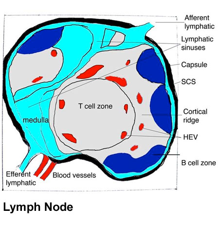 | 460 × 463 (54 KB) | Z8600021 (talk | contribs) | ==Lymph node structure== Schematic representation of the organization of a lymph node (left panel). Afferent lymphatics enter lymph nodes and deliver lymph to the subcapsular sinus (SCS), which forms a channel around the periphery of the lymph node. Lymp |
You cannot overwrite this file.
File usage
The following 4 pages use this file:


