File:HeartILP001.jpg: Difference between revisions
From Embryology
No edit summary |
No edit summary |
||
| Line 1: | Line 1: | ||
Embryo at approximately 18 days showing early angiogenesis and the development of the primordial heart tubes in the cardiogenic region. | Embryo at approximately 18 days showing early angiogenesis and the development of the primordial heart tubes in the cardiogenic region. | ||
{{Template: | {{Template:SEM}} | ||
[[category:HeartILP]] | [[category:HeartILP]] | ||
Revision as of 13:08, 8 September 2009
Embryo at approximately 18 days showing early angiogenesis and the development of the primordial heart tubes in the cardiogenic region.
Image Source: Scanning electron micrographs of the Carnegie stages of the early human embryos are reproduced with the permission of Prof Kathy Sulik, from embryos collected by Dr. Vekemans and Tania Attié-Bitach. Images are for educational purposes only and cannot be reproduced electronically or in writing without permission.
File history
Click on a date/time to view the file as it appeared at that time.
| Date/Time | Thumbnail | Dimensions | User | Comment | |
|---|---|---|---|---|---|
| current | 11:44, 8 September 2009 | 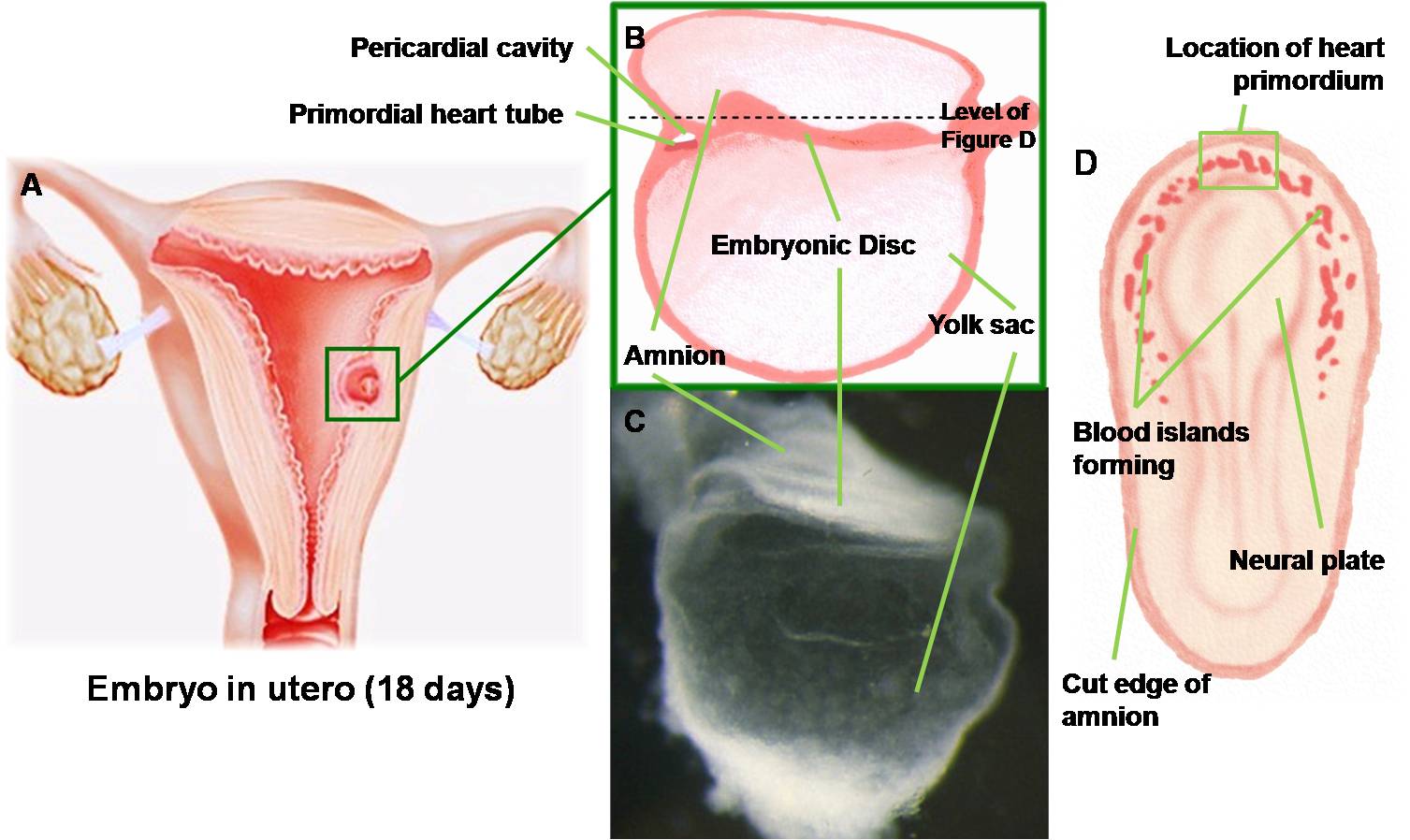 | 1,507 × 898 (125 KB) | Z3212774 (talk | contribs) | Embryo at approximately 18 days showing early angiogenesis and the development of the primordial heart tubes in the cardiogenic region. Brightfield image © Sulik. category:HeartILP |
You cannot overwrite this file.
File usage
The following page uses this file: