Talk:Lecture - Neural Development: Difference between revisions
No edit summary |
|||
| Line 1: | Line 1: | ||
==The nervous system== | |||
[[ | [[Book_-_Text-Book_of_Embryology_17|The nervous system]] | ||
<gallery> | |||
File:Bailey358.jpg|Fig. 358 | |||
File:Bailey359.jpg|Fig. 359 | |||
| | File:Bailey360.jpg|Fig. 360 | ||
| | File:Bailey361.jpg|Fig. 361 | ||
| | File:Bailey362.jpg|Fig. 362 | ||
|- | File:Bailey363.jpg|Fig. 363 | ||
| | File:Bailey364.jpg|Fig. 364 | ||
| | File:Bailey365.jpg|Fig. 365 | ||
| | File:Bailey366.jpg|Fig. 366 | ||
| | File:Bailey367.jpg|Fig. 367 | ||
File:Bailey368.jpg|Fig. 368 | |||
File:Bailey369.jpg|Fig. 369 | |||
File:Bailey370.jpg|Fig. 370 | |||
File:Bailey371.jpg|Fig. 371 | |||
File:Bailey372.jpg|Fig. 372 | |||
File:Bailey373.jpg|Fig. 373 | |||
File:Bailey374.jpg|Fig. 374 | |||
File:Bailey375.jpg|Fig. 375 | |||
File:Bailey376.jpg|Fig. 376 | |||
File:Bailey377.jpg|Fig. 377 | |||
File:Bailey378.jpg|Fig. 378 | |||
File:Bailey379-382.jpg|Fig. 379-382 | |||
File:Bailey383.jpg|Fig. 383 | |||
File:Bailey384.jpg|Fig. 384 | |||
File:Bailey385.jpg|Fig. 385 | |||
File:Bailey386.jpg|Fig. 386 | |||
File:Bailey387.jpg|Fig. 387 | |||
File:Bailey388.jpg|Fig. 388 | |||
File:Bailey389.jpg|Fig. 389 | |||
File:Bailey390.jpg|Fig. 390 | |||
File:Bailey391.jpg|Fig. 391 | |||
File:Bailey392.jpg|Fig. 392 | |||
File:Bailey393.jpg|Fig. 393 | |||
File:Bailey394.jpg|Fig. 394 | |||
File:Bailey395.jpg|Fig. 395 | |||
File:Bailey396.jpg|Fig. 396 | |||
File:Bailey397.jpg|Fig. 397 | |||
File:Bailey398.jpg|Fig. 398 | |||
File:Bailey399.jpg|Fig. 399 | |||
File:Bailey400.jpg|Fig. 400 | |||
: | File:Bailey401.jpg|Fig. 401 | ||
File:Bailey402.jpg|Fig. 402 | |||
File:Bailey403.jpg|Fig. 403 | |||
File:Bailey404.jpg|Fig. 404 | |||
File:Bailey405.jpg|Fig. 405 | |||
File:Bailey406.jpg|Fig. 406 | |||
File:Bailey407.jpg|Fig. 407 | |||
File:Bailey408.jpg|Fig. 408 | |||
File:Bailey409.jpg|Fig. 409 | |||
File:Bailey410.jpg|Fig. 410 | |||
File:Bailey411.jpg|Fig. 411 | |||
File:Bailey412.jpg|Fig. 412 | |||
File:Bailey413.jpg|Fig. 413 | |||
File:Bailey414.jpg|Fig. 414 | |||
File:Bailey415.jpg|Fig. 415 | |||
File:Bailey416.jpg|Fig. 416 | |||
File:Bailey417.jpg|Fig. 417 | |||
File:Bailey418.jpg|Fig. 418 | |||
File:Bailey419.jpg|Fig. 419 | |||
File:Bailey420.jpg|Fig. 420 | |||
File:Bailey421.jpg|Fig. 421 | |||
File:Bailey422.jpg|Fig. 422 | |||
File:Bailey423.jpg|Fig. 423 | |||
File:Bailey424.jpg|Fig. 424 | |||
File:Bailey425.jpg|Fig. 425 | |||
File:Bailey426.jpg|Fig. 426 | |||
File:Bailey427.jpg|Fig. 427 | |||
File:Bailey428.jpg|Fig. 428 | |||
File:Bailey429.jpg|Fig. 429 | |||
File:Bailey430.jpg|Fig. 430 | |||
File:Bailey431.jpg|Fig. 431 | |||
File:Bailey432.jpg|Fig. 432 | |||
File:Bailey433.jpg|Fig. 433 | |||
File:Bailey434.jpg|Fig. 434 | |||
File:Bailey435.jpg|Fig. 435 | |||
File:Bailey436.jpg|Fig. 436 | |||
File:Bailey437.jpg|Fig. 437 | |||
File:Bailey438.jpg|Fig. 438 | |||
File:Bailey439.jpg|Fig. 439 | |||
File:Bailey440.jpg|Fig. 440 | |||
File:Bailey441.jpg|Fig. 441 | |||
File:Bailey442.jpg|Fig. 442 | |||
File:Bailey443.jpg|Fig. 443 | |||
File:Bailey444.jpg|Fig. 444 | |||
File:Bailey445.jpg|Fig. 445 | |||
File:Bailey446.jpg|Fig. 446 | |||
File:Bailey447.jpg|Fig. 447 | |||
File:Bailey448.jpg|Fig. 448 | |||
File:Bailey449.jpg|Fig. 449 | |||
File:Bailey450.jpg|Fig. 450 | |||
File:Bailey451.jpg|Fig. 451 | |||
File:Bailey452.jpg|Fig. 452 | |||
File:Bailey453.jpg|Fig. 453 | |||
File:Bailey454.jpg|Fig. 454 | |||
File:Bailey455.jpg|Fig. 455 | |||
</gallery> | |||
Revision as of 11:42, 19 August 2011
The nervous system
Fig. 358
Fig. 359
Fig. 360

Fig. 361
Fig. 362
Fig. 363
Fig. 364
Fig. 365
Fig. 366
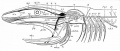
Fig. 367
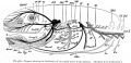
Fig. 368
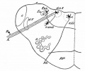
Fig. 369
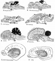
Fig. 370
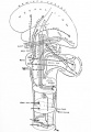
Fig. 371
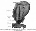
Fig. 372
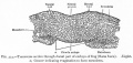
Fig. 373
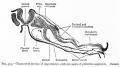
Fig. 374
Fig. 375
Fig. 376
Fig. 377
Fig. 378
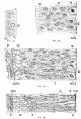
Fig. 379-382
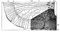
Fig. 383
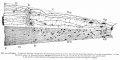
Fig. 384
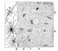
Fig. 385
Fig. 386
Fig. 387
Fig. 388
Fig. 389
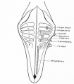
Fig. 390
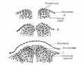
Fig. 391
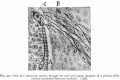
Fig. 392
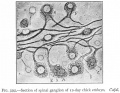
Fig. 393
Fig. 394
Fig. 395
Fig. 396
Fig. 397
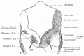
Fig. 398
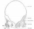
Fig. 399
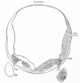
Fig. 400
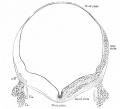
Fig. 401
Fig. 402
Fig. 403
Fig. 404
Fig. 405

Fig. 406
Fig. 407
Fig. 408
Fig. 409
Fig. 410
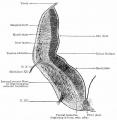
Fig. 411
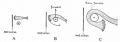
Fig. 412
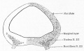
Fig. 413
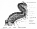
Fig. 414
Fig. 415
Fig. 416
Fig. 417
Fig. 418
Fig. 419
Fig. 420
Fig. 421
Fig. 422
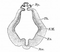
Fig. 423
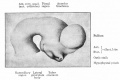
Fig. 424
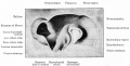
Fig. 425
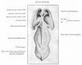
Fig. 426
Fig. 427
Fig. 428
Fig. 429
Fig. 430
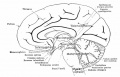
Fig. 431
Fig. 432
Fig. 433
Fig. 434
Fig. 435
Fig. 436
Fig. 437
Fig. 438
Fig. 439
Fig. 440
Fig. 441
Fig. 442
Fig. 443
Fig. 444
Fig. 445
Fig. 446
Fig. 447
Fig. 448
Fig. 449
Fig. 450
- Bailey451.jpg
Fig. 451
- Bailey452.jpg
Fig. 452
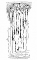
Fig. 453
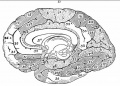
Fig. 454
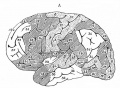
Fig. 455


