Talk:BGDA Practical 3 - Gametogenesis: Difference between revisions
(Created page with "===2010 Practical Audio=== {| border='0px' |- | 60px|left | BGD Cycle A 2010 Audio - Dr Mark Hill Monday 12th May 2010 12-2pm G2G4. :Note - this is a l...") |
No edit summary |
||
| Line 11: | Line 11: | ||
[[Media:BGD2010-Embryo Lab 120510-304.mp3|listen Part 4]] | [[:File:BGD2010-Embryo Lab 120510-304.mp3|download]] (1.2 Mb MP3 9:29) | [[Media:BGD2010-Embryo Lab 120510-304.mp3|listen Part 4]] | [[:File:BGD2010-Embryo Lab 120510-304.mp3|download]] (1.2 Mb MP3 9:29) | ||
|- | |||
|} | |||
==Meiosis Sex Differences== | |||
'''Female''' (oogenesis) | |||
* Meiosis initiated once in a finite population of cells | |||
* 1 gamete produced / meiosis | |||
* Completion of meiosis delayed for months or years | |||
* Meiosis arrested at 1st meiotic prophase and reinitiated in a smaller population of cells | |||
* Differentiation of gamete occurs while diploid in first meiotic prophase | |||
* All chromosomes exhibit equivalent transcription and recombination during meiotic prophase | |||
'''Male''' (spermatogenesis) | |||
* Meiosis initiated continuously in a mitotically dividing stem cell population | |||
* 4 gametes produced / meiosis | |||
* Meiosis completed in days or weeks | |||
* Meiosis and differentiation proceed continuously without cell cycle arrest | |||
* Differentiation of gamete occurs while haploid after meiosis ends | |||
Sex chromosomes excluded from recombination and transcription during first meiotic prophase | |||
== Female Gametogenesis == | |||
In females, the total number of eggs ever to be produced are present in the newborn female. | |||
# All eggs are arrested at an early stage of the first meiotic division as a primary oocyte (primordial follicle). Following purberty, during each menstrual cycle, pituitary gonadotrophin stimulates completion of '''meiosis 1''' the day before ovulation. | |||
# In '''meiosis 1''', a diploid cell becomes 2 haploid (23 chromosomes) daughter cells, each chromosome has two chromatids. One cell becomes the secondary oocyte the other cell forms the first polar body. | |||
# The secondary oocyte then commences '''meiosis 2''' which arrests at metaphase and will not continue without fertilization. | |||
# At fertilization '''meiosis 2''' completes, forming a second polar body. Note that the first polar body may also undergo this process forming a third polar body. | |||
[[File:Female_gametogenesis.jpg|600px|Female gametogenesis]] | |||
===Polar Body=== | |||
[[File:Human oocyte-metaphase II.jpg|thumb|Human oocyte at metaphase II showing polar body at 12 o'clock position.]] | |||
The breakdown of the germinal vesicle indicates a resumption of meiosis and the extrusion of the first polar body (1 PB) indicates completion of the first meiotic division in human oocytes. The polar body is a small cytoplasmic exclusion body formed to enclose the excess DNA formed during the oocyte (egg) meiosis and following sperm fertilization. There are 2-3 polar bodies derived from the oocyte present in the zygote, the number is dependent upon whether polar body 1 (the first polar body formed during meiosis 1) divides during meiosis 2. This exclusion body contains the excess DNA from the reductive division (the second and third polar bodies are formed from meiosis 2 at fertilization). These polar bodies do not contribute to the future genetic complement of the zygote, embryo or fetus. | |||
Recent research in some species suggest that the space formed by the peripheral polar body (between the oocyte and the zona pellucia) can influence the site of spermatozoa fertilization. | |||
Assisted reproductive techniques involving intracytoplasmic sperm injection (ICSI) have looked at the "quality" of the polar body and found that the morphology is related to mature oocyte viability and has the potential to predict oocyte fertilization rates and pregnancy achievement.<ref><pubmed>10655316</pubmed></ref><ref><pubmed>19960239</pubmed>| [http://www.ncbi.nlm.nih.gov/pmc/articles/PMC2799563 PMC2799563]</ref> | |||
===Female Abnormalities=== | |||
[[File:Trisomy21female.jpg|thumb|Trisomy 21 female karyotype]] | |||
Meiotic non-disjunction resulting in aneuploidy, most are embryonic lethal and not seen. The potential for genetic abnormalities increase with maternal age. | |||
* Autosomal chromosome aneuploidy | |||
** trisomy 21 - [[trisomy 21|Down syndrome]] | |||
** trisomy 18 - Edwards syndrome | |||
** trisomy 13 - Patau syndrome | |||
* Sex chromosome aneuploidy | |||
** monosomy X - Turner's Syndrome | |||
** trisomy X - Triple-X syndrome | |||
** 47 XXY - Klinefelter's Syndrome | |||
== Male Gametogenesis == | |||
In males, sperm continues to be generated throughout life from a stem cell population in the testis. Spermatozoa maturation involves two processes meiosis and spermiogenesis | |||
[[File:Male_gametogenesis.jpg|600px]] | |||
The above figure compares meiosis to the female (the polar bodies have been removed and labelling updated). | |||
<gallery> | |||
File:Historic-testis.jpg|Historic testis drawing | |||
File:Seminiferous-tubule-HEx40.jpg|Adult Seminiferous tubule showing spermatozoa developmental stages | |||
File:Testis_histology_2.jpg|Seminiferous tubule cross-section and supporting cells | |||
File:Human-spermatozoa.jpg|Human spermatozoa | |||
</gallery> | |||
===Human Spermatozoa Development=== | |||
* Spermatogenesis process of spermatagonia mature into spermatazoa (sperm). | |||
* Continuously throughout life occurs in the seminiferous tubules in the male gonad- testis (plural testes). | |||
* At puberty spermatagonia activate and proliferate (mitosis). | |||
* about 48 days from entering meiosis until morphologically mature spermatozoa | |||
* about 64 days to complete spermatogenesis, depending reproduction time of spermatogonia | |||
* follicle stimulating hormone (FSH) - stimulates the spermatogenic epithelium | |||
* luteinizing-hormone (LH) - stimulates testosterone production by Leydig cells | |||
[[File:Human-spermatozoa_EM01.jpg|600px]] | |||
{| | |||
|- | |||
| [[File:Spermatozoa animation icon.jpg|200px|link=Development_Animation_-_Spermatozoa]] | |||
| '''Mature human spermatozoa''' | |||
* 60 µm long, actively motile | |||
* divided into 3 main regions (head, neck and tail) | |||
* head - (flattened, 5 µm long by 3 µm wide) the nucleus and acrosome. Posterior part of nuclear membrane forms the basal plate. | |||
* neck - (1 µm) attached to basal plate, transverse oriented centriole, contains nine segmented columns of fibrous material, continue as outer dense fibres in tail. | |||
* tail - 3 parts a middle piece, principal piece and end piece | |||
** middle piece - (5 µm long) [[A#axonema|axonema]] and dense fibres surrounded by mitochondria | |||
** principal piece - (45 µm long) fibrous sheath interconnected by regularly spaced circumferential hoops | |||
** end piece - (5 µm long) [[A#axonema|axonema]] surrounded by small amount of cytoplasm and plasma membrane | |||
|- | |- | ||
|} | |} | ||
Revision as of 13:41, 8 May 2011
2010 Practical Audio
 |
BGD Cycle A 2010 Audio - Dr Mark Hill Monday 12th May 2010 12-2pm G2G4.
listen Part 4 | download (1.2 Mb MP3 9:29) |
Meiosis Sex Differences
Female (oogenesis)
- Meiosis initiated once in a finite population of cells
- 1 gamete produced / meiosis
- Completion of meiosis delayed for months or years
- Meiosis arrested at 1st meiotic prophase and reinitiated in a smaller population of cells
- Differentiation of gamete occurs while diploid in first meiotic prophase
- All chromosomes exhibit equivalent transcription and recombination during meiotic prophase
Male (spermatogenesis)
- Meiosis initiated continuously in a mitotically dividing stem cell population
- 4 gametes produced / meiosis
- Meiosis completed in days or weeks
- Meiosis and differentiation proceed continuously without cell cycle arrest
- Differentiation of gamete occurs while haploid after meiosis ends
Sex chromosomes excluded from recombination and transcription during first meiotic prophase
Female Gametogenesis
In females, the total number of eggs ever to be produced are present in the newborn female.
- All eggs are arrested at an early stage of the first meiotic division as a primary oocyte (primordial follicle). Following purberty, during each menstrual cycle, pituitary gonadotrophin stimulates completion of meiosis 1 the day before ovulation.
- In meiosis 1, a diploid cell becomes 2 haploid (23 chromosomes) daughter cells, each chromosome has two chromatids. One cell becomes the secondary oocyte the other cell forms the first polar body.
- The secondary oocyte then commences meiosis 2 which arrests at metaphase and will not continue without fertilization.
- At fertilization meiosis 2 completes, forming a second polar body. Note that the first polar body may also undergo this process forming a third polar body.

Polar Body

The breakdown of the germinal vesicle indicates a resumption of meiosis and the extrusion of the first polar body (1 PB) indicates completion of the first meiotic division in human oocytes. The polar body is a small cytoplasmic exclusion body formed to enclose the excess DNA formed during the oocyte (egg) meiosis and following sperm fertilization. There are 2-3 polar bodies derived from the oocyte present in the zygote, the number is dependent upon whether polar body 1 (the first polar body formed during meiosis 1) divides during meiosis 2. This exclusion body contains the excess DNA from the reductive division (the second and third polar bodies are formed from meiosis 2 at fertilization). These polar bodies do not contribute to the future genetic complement of the zygote, embryo or fetus.
Recent research in some species suggest that the space formed by the peripheral polar body (between the oocyte and the zona pellucia) can influence the site of spermatozoa fertilization.
Assisted reproductive techniques involving intracytoplasmic sperm injection (ICSI) have looked at the "quality" of the polar body and found that the morphology is related to mature oocyte viability and has the potential to predict oocyte fertilization rates and pregnancy achievement.[1][2]
Female Abnormalities

Meiotic non-disjunction resulting in aneuploidy, most are embryonic lethal and not seen. The potential for genetic abnormalities increase with maternal age.
- Autosomal chromosome aneuploidy
- trisomy 21 - Down syndrome
- trisomy 18 - Edwards syndrome
- trisomy 13 - Patau syndrome
- Sex chromosome aneuploidy
- monosomy X - Turner's Syndrome
- trisomy X - Triple-X syndrome
- 47 XXY - Klinefelter's Syndrome
Male Gametogenesis
In males, sperm continues to be generated throughout life from a stem cell population in the testis. Spermatozoa maturation involves two processes meiosis and spermiogenesis

The above figure compares meiosis to the female (the polar bodies have been removed and labelling updated).
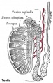
Historic testis drawing

Adult Seminiferous tubule showing spermatozoa developmental stages

Seminiferous tubule cross-section and supporting cells
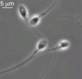
Human spermatozoa


Human Spermatozoa Development
- Spermatogenesis process of spermatagonia mature into spermatazoa (sperm).
- Continuously throughout life occurs in the seminiferous tubules in the male gonad- testis (plural testes).
- At puberty spermatagonia activate and proliferate (mitosis).
- about 48 days from entering meiosis until morphologically mature spermatozoa
- about 64 days to complete spermatogenesis, depending reproduction time of spermatogonia
- follicle stimulating hormone (FSH) - stimulates the spermatogenic epithelium
- luteinizing-hormone (LH) - stimulates testosterone production by Leydig cells

Mature human spermatozoa
|
- ↑ <pubmed>10655316</pubmed>
- ↑ <pubmed>19960239</pubmed>| PMC2799563