File:HamiltonBoyd1960 fig07.jpg: Difference between revisions
mNo edit summary |
mNo edit summary |
||
| Line 3: | Line 3: | ||
Note that the uterine lumen is still widely patent. A section through the uterus and in situ placenta of this specimen is illustrated in Pl. 9, fig. 25. | Note that the uterine lumen is still widely patent. A section through the uterus and in situ placenta of this specimen is illustrated in Pl. 9, fig. 25. | ||
{{HamiltonBoyd1960 plates footer}} | |||
Latest revision as of 18:40, 6 August 2020
Fig. 7.
Photograph ( x 0-9) of a coronal section of a uterus containing a 29 mm. embryo (CX. 105).
Note that the uterine lumen is still widely patent. A section through the uterus and in situ placenta of this specimen is illustrated in Pl. 9, fig. 25.
Plates: 1 | 2 | 3 | 4 | 5 | 6 | 7 | 8 | 9 | 10 | 11 | 12 | 13
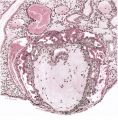
Plate 1
Plate 2
Plate 3
Plate 4
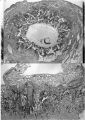
Plate 5
Plate 6
Plate 7
Plate 8
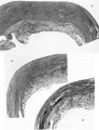
Plate 9
Plate 10
Plate 11
Plate 12
Plate 13


Reference
Hamilton WJ. and Boyd JD. Development of the human placenta in the first three months of gestation. (1960) J Anat. 94(3): 297-328. PMID14399291 | PDF
Cite this page: Hill, M.A. (2024, June 27) Embryology HamiltonBoyd1960 fig07.jpg. Retrieved from https://embryology.med.unsw.edu.au/embryology/index.php/File:HamiltonBoyd1960_fig07.jpg
- © Dr Mark Hill 2024, UNSW Embryology ISBN: 978 0 7334 2609 4 - UNSW CRICOS Provider Code No. 00098G
File history
Yi efo/eka'e gwa ebo wo le nyangagi wuncin ye kamina wunga tinya nan
| Gwalagizhi | Nyangagi | Dimensions | User | Comment | |
|---|---|---|---|---|---|
| current | 18:36, 6 August 2020 | 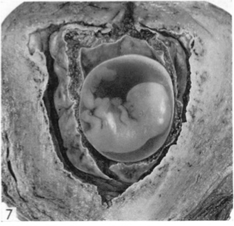 | 800 × 775 (122 KB) | Z8600021 (talk | contribs) |
You cannot overwrite this file.
File usage
The following 2 pages use this file: