File:Johnston1907 fig015.jpg: Difference between revisions
From Embryology
mNo edit summary |
(Z8600021 uploaded a new version of File:Johnston1907 fig015.jpg) |
(No difference)
| |
Revision as of 22:11, 23 February 2020
Summary
Fig 15. Transverse section through neural tube, neural crest and ectoderm of Amblystoma punctatum at a later stage than that shown in Fig. 14.
Johnston JB. The Nervous System of Vertebrates. (1907) Blakiston's Son & Co., London.
| Historic Disclaimer - information about historic embryology pages |
|---|
 |
Cite this page: Hill, M.A. (2024, June 14) Embryology Johnston1907 fig015.jpg. Retrieved from https://embryology.med.unsw.edu.au/embryology/index.php/File:Johnston1907_fig015.jpg
- © Dr Mark Hill 2024, UNSW Embryology ISBN: 978 0 7334 2609 4 - UNSW CRICOS Provider Code No. 00098G
File history
Click on a date/time to view the file as it appeared at that time.
| Date/Time | Thumbnail | Dimensions | User | Comment | |
|---|---|---|---|---|---|
| current | 22:11, 23 February 2020 |  | 800 × 703 (100 KB) | Z8600021 (talk | contribs) | Reverted to version as of 23:06, 23 February 2020 (AEDT) |
| 22:11, 23 February 2020 | 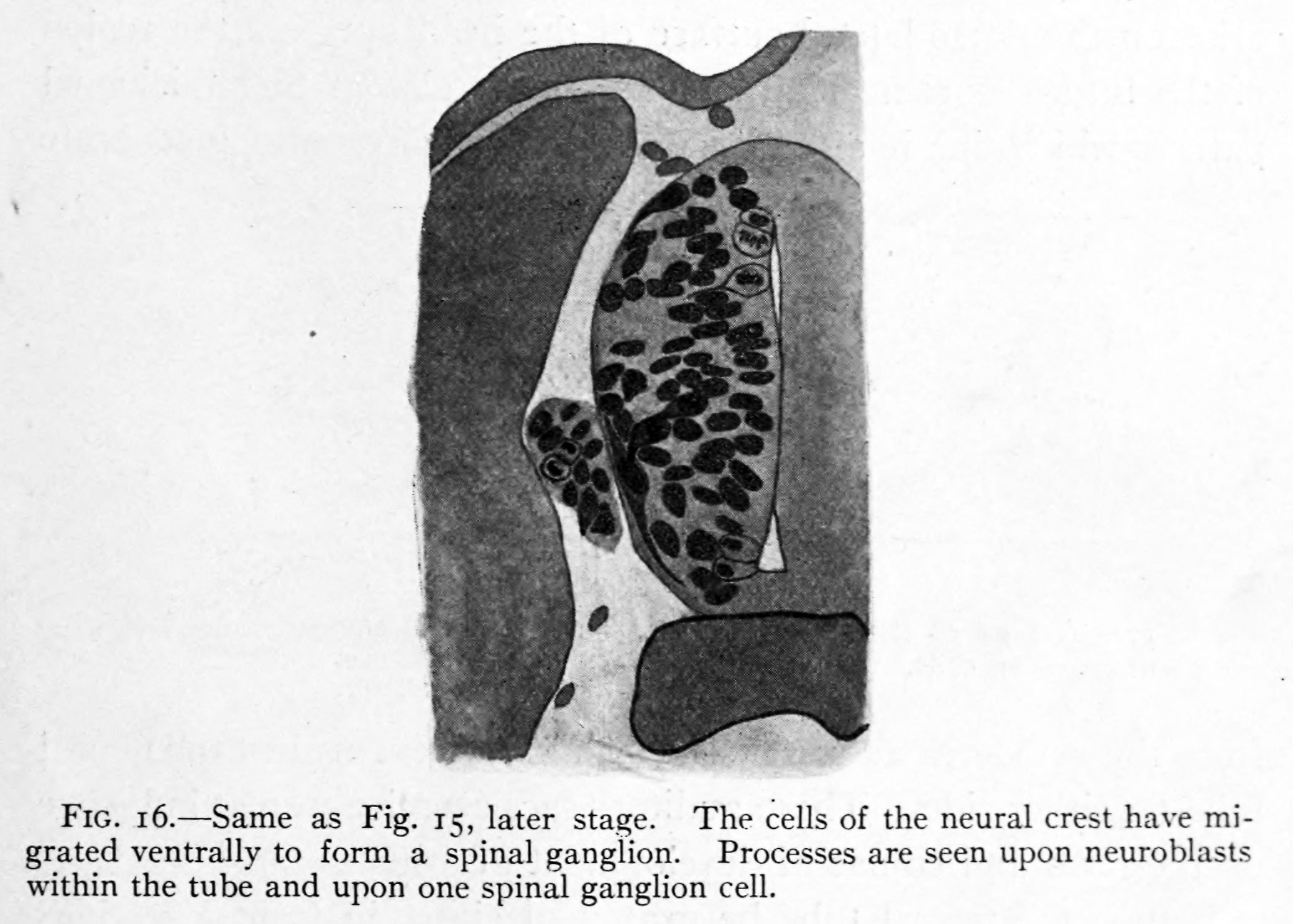 | 2,080 × 1,487 (331 KB) | Z8600021 (talk | contribs) | ||
| 22:06, 23 February 2020 | 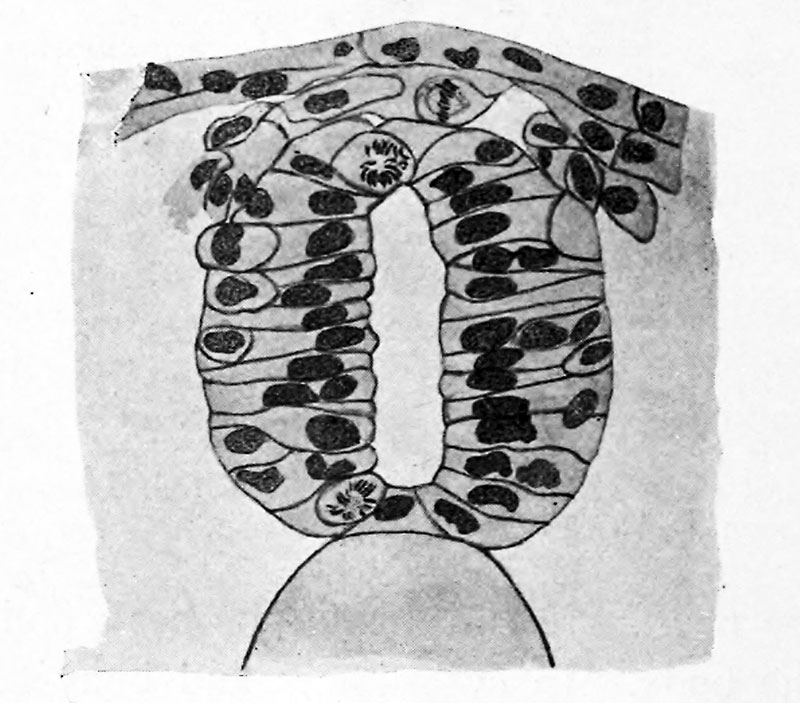 | 800 × 703 (100 KB) | Z8600021 (talk | contribs) | ||
| 22:04, 23 February 2020 | 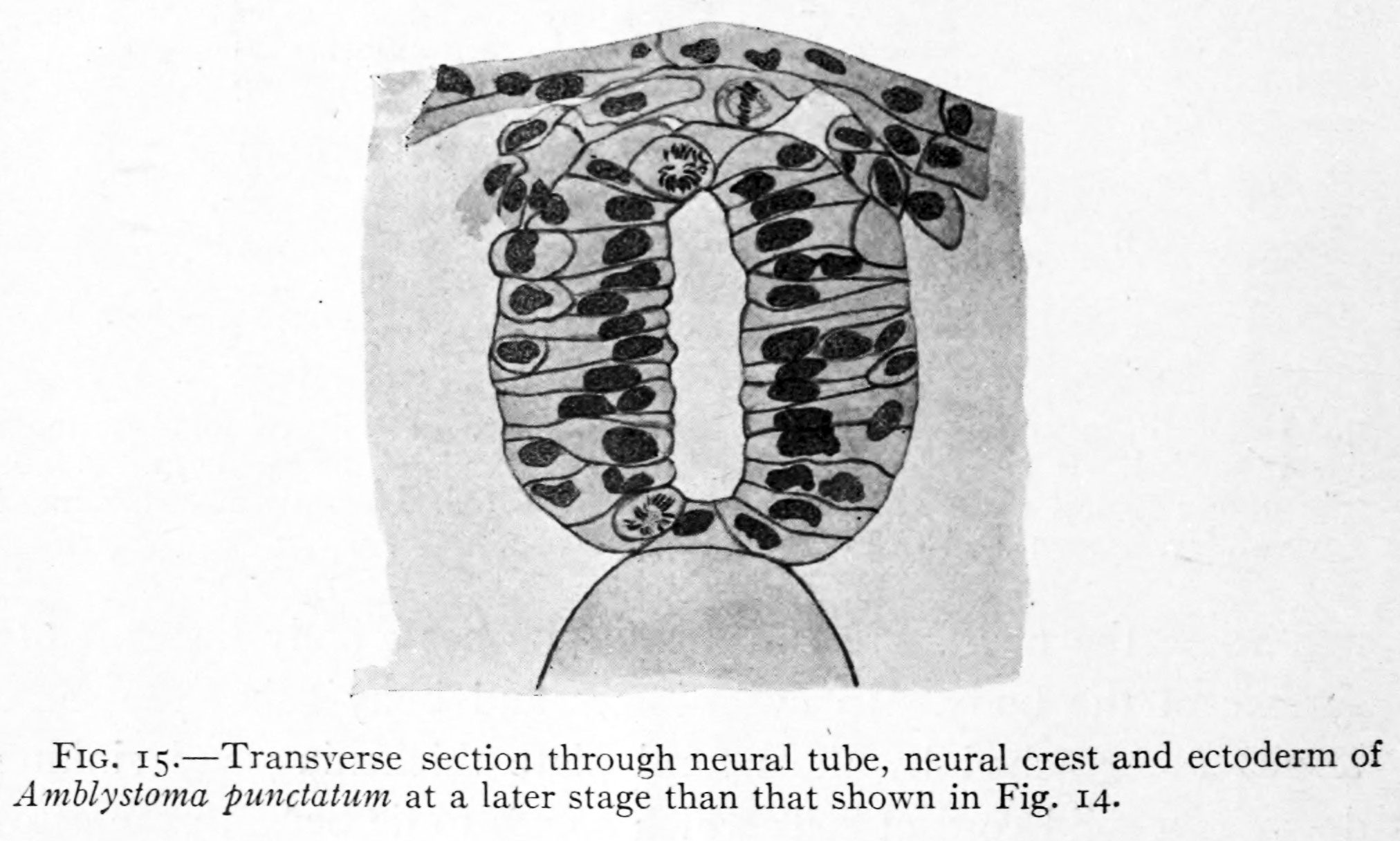 | 2,024 × 1,216 (247 KB) | Z8600021 (talk | contribs) | Fig 15. Transverse section through neural tube, neural crest and ectoderm of Amblystoma punctatum at a later stage than that shown in Fig. 14. |
You cannot overwrite this file.
File usage
The following 2 pages use this file: