File:Cullen1916 fig28.jpg: Difference between revisions
(Z8600021 uploaded a new version of File:Cullen1916 fig28.jpg) |
mNo edit summary |
||
| Line 1: | Line 1: | ||
Fig. 28. | ==Fig. 28. The Umbilical Region in a Fetus about Five Months Old Viewed from the Left== | ||
Note the portion of the triangular falciform Ligament between the umbilical vein and the ventral abdominal wall. The bladder and allantois (urachus) are in the usual position; the latter is becoming obliterated. In the cord it appears below and between the umbilical arteries. In the center of the cord is the sickle-shaped lumen of the exoccelom, which, however, is bridged over in many places by delicate strands of connective tissue. In the abdomen a shallow pit still marks the position of the umbilical ring. | |||
{{Cullen1916 figures1}} | |||
Latest revision as of 14:16, 28 October 2018
Fig. 28. The Umbilical Region in a Fetus about Five Months Old Viewed from the Left
Note the portion of the triangular falciform Ligament between the umbilical vein and the ventral abdominal wall. The bladder and allantois (urachus) are in the usual position; the latter is becoming obliterated. In the cord it appears below and between the umbilical arteries. In the center of the cord is the sickle-shaped lumen of the exoccelom, which, however, is bridged over in many places by delicate strands of connective tissue. In the abdomen a shallow pit still marks the position of the umbilical ring.
| Historic Disclaimer - information about historic embryology pages |
|---|
 |
- Figure Links: 1 Human embryo 0.7 mm | 2 Human embryo 1.7 mm | 3 Human embryo 2.5 mm | 4 Human embryo 3.5 mm | 5 Human embryo 5 mm | 6 Human embryo 7 mm | 7 Human embryo 7 mm | 8 Human embryo 10 mm | 9 Human embryo 12.5 mm | 10 Human embryo 10 mm | 11 Human embryo 23 mm | 12 Human embryo 3 cm | 13 Human embryo 4.5 cm sagittal | 14 Human Embryo 4.5 cm | 15 Human Embryo 5.2 cm | 16 Human Embryo 6.5 cm | 17 Human Embryo 7.5 cm | 18 Human Embryo 9 cm | 19 Human Embryo 10 cm | 20 Human Embryo 12 cm | 21 Human Embryo 12 cm | 22 Human Embryo 12 cm | 23 Human Embryo 12 cm Cord | 28 Fetus Five Months | 30 Ventral Heria | 31 Human Embryo 5.5 cm | 32 Term Human | 33 Term Human | [[Figures
Reference
Cullen TS. Embryology, anatomy, and diseases of the umbilicus together with diseases of the urachus. (1916) W. B. Saunders Company, Philadelphia And London.
Cite this page: Hill, M.A. (2024, June 27) Embryology Cullen1916 fig28.jpg. Retrieved from https://embryology.med.unsw.edu.au/embryology/index.php/File:Cullen1916_fig28.jpg
- © Dr Mark Hill 2024, UNSW Embryology ISBN: 978 0 7334 2609 4 - UNSW CRICOS Provider Code No. 00098G
File history
Yi efo/eka'e gwa ebo wo le nyangagi wuncin ye kamina wunga tinya nan
| Gwalagizhi | Nyangagi | Dimensions | User | Comment | |
|---|---|---|---|---|---|
| current | 14:14, 28 October 2018 | 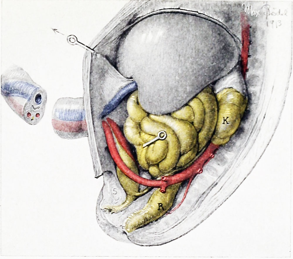 | 1,000 × 882 (141 KB) | Z8600021 (talk | contribs) | |
| 14:13, 28 October 2018 | 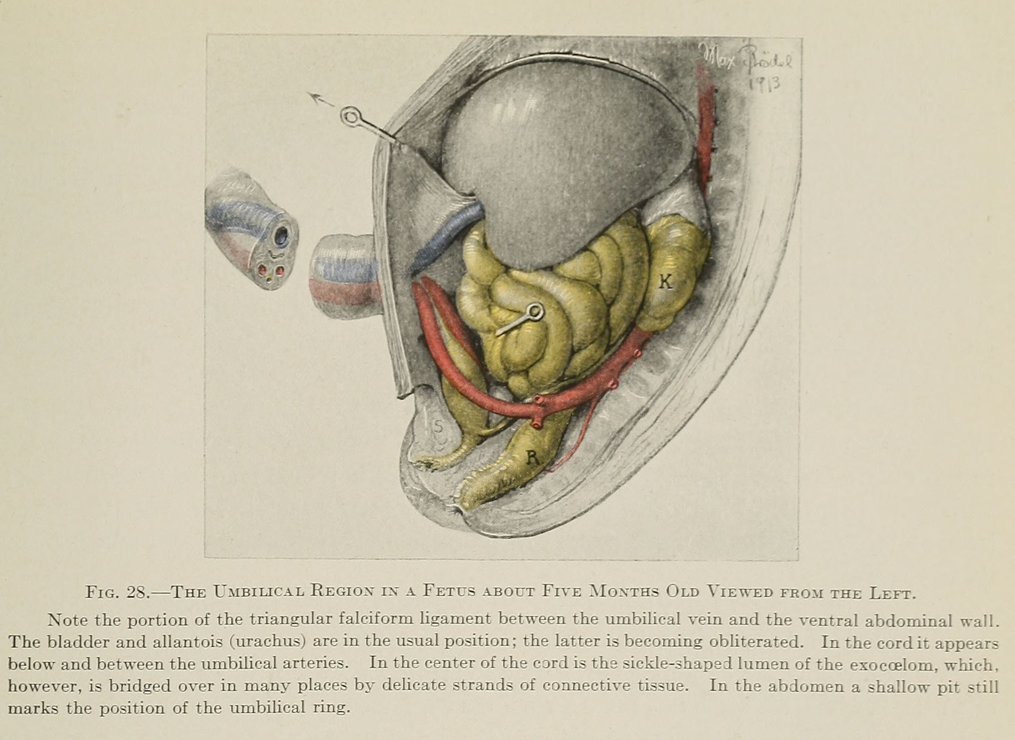 | 2,045 × 1,492 (362 KB) | Z8600021 (talk | contribs) | Fig. 28. — The Umbilical Region in a Fetus about Five Months Old Viewed from the Left. Note the portion of the triangular falciform Ligament between the umbilical vein and the ventral abdominal wall. The bladder and allantois (urachus) are in the usu... |
You cannot overwrite this file.
File usage
The following 2 pages use this file: