File:Hamilton1943 plate01.jpg: Difference between revisions
(Z8600021 uploaded a new version of File:Hamilton1943 plate01.jpg) |
mNo edit summary |
||
| Line 1: | Line 1: | ||
==Plate 1== | |||
Fig. 1. Photomicrograph of ovum No. 1 in the living state. | |||
Fig. 2. The same ovum immediately after fixation. | |||
Fig. 3. A section of ovum No. 1 showing the metaphase of The dark areas on the vitellus are corona radiata cells on the the second maturation division. The first polar body (out upper or lower surface of the zona. x .180. of focus in the upper right—hand corner) is seen. The vitellns has shrunk from the zona pellucida and most of the corona radiata cells have been lost. (cf. P]. I, fig. I). x820. | |||
Fig. 4. Photomicrograph of ovum No. 2. A con} lete sperm is seen x 480. in the zona pellucida: other sperm heads are visiblle. At the upper right-hand corner of the photograph globular detritus is seen. x 480. w..H. | |||
Fig. 5. A surface view of the endometrium of specimen No. 3. The smooth elevation produced by the implanting embryo is seen. The endometritnn shows fissures :md crct'iccs which for the most part are associated with the mouths of the uterine glands. x 15. | |||
===Reference=== | ===Reference=== | ||
{{Ref-Hamilton1943}} | {{Ref-Hamilton1943}} | ||
Revision as of 20:39, 30 October 2017
Plate 1
Fig. 1. Photomicrograph of ovum No. 1 in the living state.
Fig. 2. The same ovum immediately after fixation.
Fig. 3. A section of ovum No. 1 showing the metaphase of The dark areas on the vitellus are corona radiata cells on the the second maturation division. The first polar body (out upper or lower surface of the zona. x .180. of focus in the upper right—hand corner) is seen. The vitellns has shrunk from the zona pellucida and most of the corona radiata cells have been lost. (cf. P]. I, fig. I). x820.
Fig. 4. Photomicrograph of ovum No. 2. A con} lete sperm is seen x 480. in the zona pellucida: other sperm heads are visiblle. At the upper right-hand corner of the photograph globular detritus is seen. x 480. w..H.
Fig. 5. A surface view of the endometrium of specimen No. 3. The smooth elevation produced by the implanting embryo is seen. The endometritnn shows fissures :md crct'iccs which for the most part are associated with the mouths of the uterine glands. x 15.
Reference
Hamilton WJ. Barnes J. and Dodds GH. Phases of maturation, fertilization and early development in man. (1943) J. Obstet. Gynaecol, Brit. Emp., 50: 241-245.
File history
Yi efo/eka'e gwa ebo wo le nyangagi wuncin ye kamina wunga tinya nan
| Gwalagizhi | Nyangagi | Dimensions | User | Comment | |
|---|---|---|---|---|---|
| current | 20:36, 30 October 2017 |  | 1,944 × 2,644 (387 KB) | Z8600021 (talk | contribs) | |
| 20:35, 30 October 2017 | 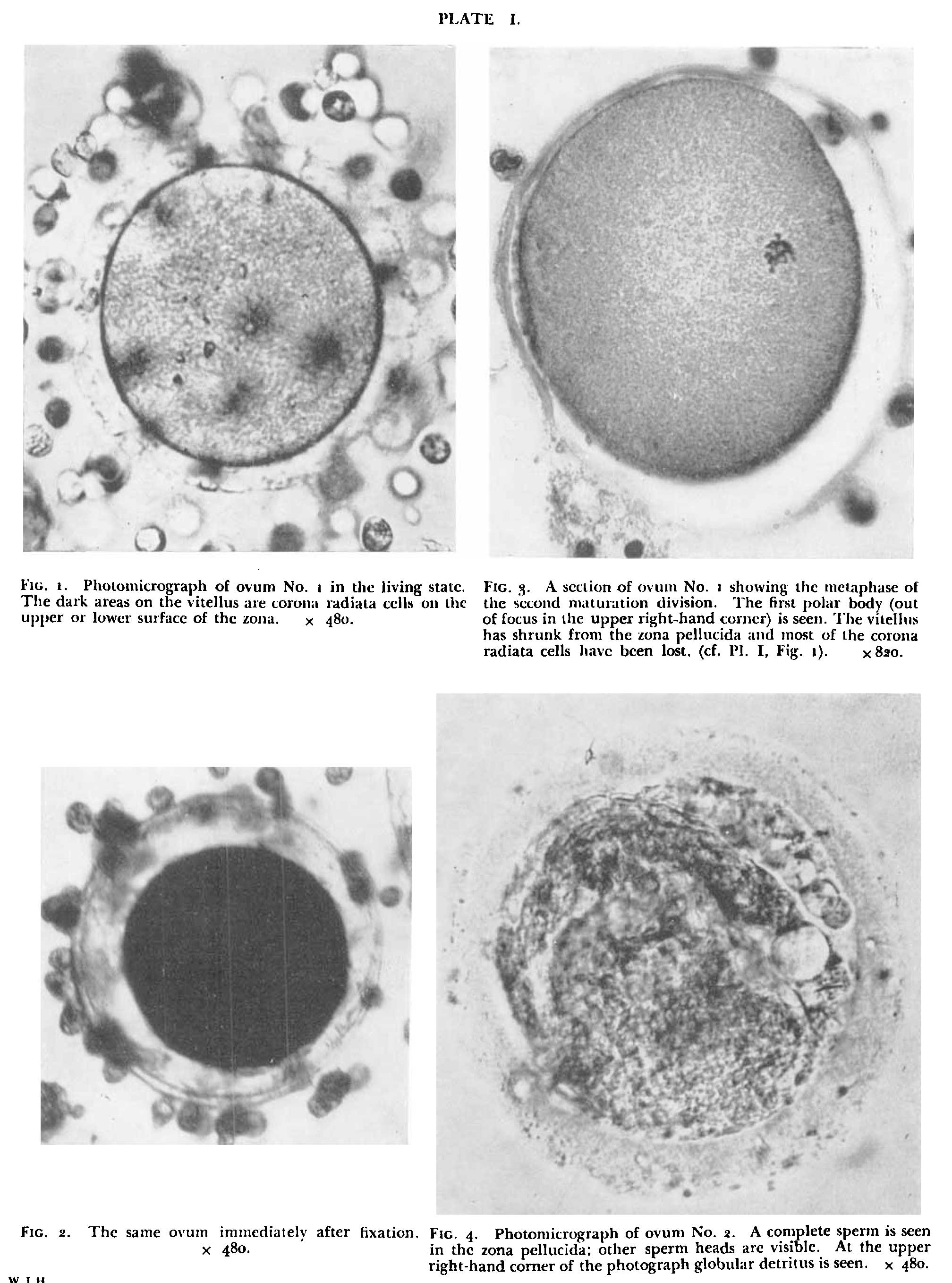 | 2,016 × 2,716 (370 KB) | Z8600021 (talk | contribs) | ===Reference=== {{Ref-Hamilton1943}} |
You cannot overwrite this file.
File usage
The following page uses this file: