File:Haines1947 fig01.jpg: Difference between revisions
From Embryology
No edit summary |
mNo edit summary |
||
| Line 1: | Line 1: | ||
==Fig. 1. 10 mm Lucas Keene’s ‘2387’, 50.4. Fore-limb== | |||
The skeletal blastema (sIc.) shows clearly in the humeral, elbow (el.) and fore-arm regions, while distally it fades away towards the marginal vein. The pre-muscle masses of the upper arm on both flexor (fl.) and extensor (e:c.) aspects, the brachial plexus ( pl.) and median nerve (md.) are visible. A blood vessel of the interosseous group (b.v.) pierces the blastema. | |||
===Reference=== | |||
{{Ref-Haines1947}} | |||
{{Footer}} | |||
[[Category:Joint]][[Category:1940's]] | |||
Latest revision as of 15:23, 3 October 2017
Fig. 1. 10 mm Lucas Keene’s ‘2387’, 50.4. Fore-limb
The skeletal blastema (sIc.) shows clearly in the humeral, elbow (el.) and fore-arm regions, while distally it fades away towards the marginal vein. The pre-muscle masses of the upper arm on both flexor (fl.) and extensor (e:c.) aspects, the brachial plexus ( pl.) and median nerve (md.) are visible. A blood vessel of the interosseous group (b.v.) pierces the blastema.
Reference
Haines RW. The development of joints. (1947) J. Anat. 81, 33-55.
Cite this page: Hill, M.A. (2024, June 14) Embryology Haines1947 fig01.jpg. Retrieved from https://embryology.med.unsw.edu.au/embryology/index.php/File:Haines1947_fig01.jpg
- © Dr Mark Hill 2024, UNSW Embryology ISBN: 978 0 7334 2609 4 - UNSW CRICOS Provider Code No. 00098G
File history
Click on a date/time to view the file as it appeared at that time.
| Date/Time | Thumbnail | Dimensions | User | Comment | |
|---|---|---|---|---|---|
| current | 15:21, 3 October 2017 | 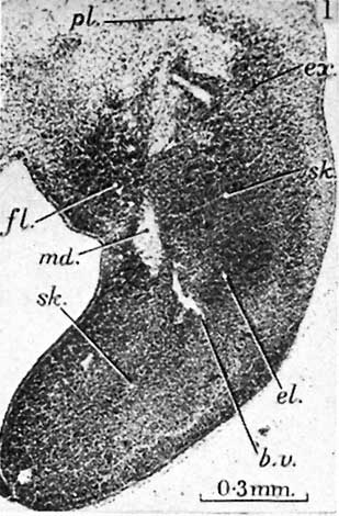 | 309 × 470 (37 KB) | Z8600021 (talk | contribs) |
You cannot overwrite this file.
File usage
The following page uses this file: