File:Hertig1946b fig09a.jpg: Difference between revisions
mNo edit summary |
mNo edit summary |
||
| Line 1: | Line 1: | ||
==Fig. 9. A. A drawing of the right half of a 2.5 mm embryo== | ==Fig. 9.A. A drawing of the right half of a 2.5 mm embryo== | ||
Representing a stage in the middle of the 4th week of development. The circulation is complete and the heart functions at this stage. Note the constriction of the yolk-sac to form the omphalomesenteric or vitelline duct as the digestive tract becomes more mature. The growth of the embryo and its surrounding amnion have resulted in the partial approximation of the body stalk and the vitelline duct so that these structures are being combined to form an early umbilical cord. (Fig. 3 from Cullen’s “The Umbilicus and Its Diseases,” W. B. Saunders Company.) | Representing a stage in the middle of the 4th week of development. The circulation is complete and the heart functions at this stage. Note the constriction of the yolk-sac to form the omphalomesenteric or vitelline duct as the digestive tract becomes more mature. The growth of the embryo and its surrounding amnion have resulted in the partial approximation of the body stalk and the vitelline duct so that these structures are being combined to form an early umbilical cord. (Fig. 3 from Cullen’s “The Umbilicus and Its Diseases,” W. B. Saunders Company.) | ||
Latest revision as of 17:35, 7 August 2017
Fig. 9.A. A drawing of the right half of a 2.5 mm embryo
Representing a stage in the middle of the 4th week of development. The circulation is complete and the heart functions at this stage. Note the constriction of the yolk-sac to form the omphalomesenteric or vitelline duct as the digestive tract becomes more mature. The growth of the embryo and its surrounding amnion have resulted in the partial approximation of the body stalk and the vitelline duct so that these structures are being combined to form an early umbilical cord. (Fig. 3 from Cullen’s “The Umbilicus and Its Diseases,” W. B. Saunders Company.)
References
Hertig AT. lnvolution of tissues in fetal life: a review. (1946) Anat. Rec. 94: 96-116.
Cite this page: Hill, M.A. (2024, June 24) Embryology Hertig1946b fig09a.jpg. Retrieved from https://embryology.med.unsw.edu.au/embryology/index.php/File:Hertig1946b_fig09a.jpg
- © Dr Mark Hill 2024, UNSW Embryology ISBN: 978 0 7334 2609 4 - UNSW CRICOS Provider Code No. 00098G
File history
Yi efo/eka'e gwa ebo wo le nyangagi wuncin ye kamina wunga tinya nan
| Gwalagizhi | Nyangagi | Dimensions | User | Comment | |
|---|---|---|---|---|---|
| current | 17:34, 7 August 2017 | 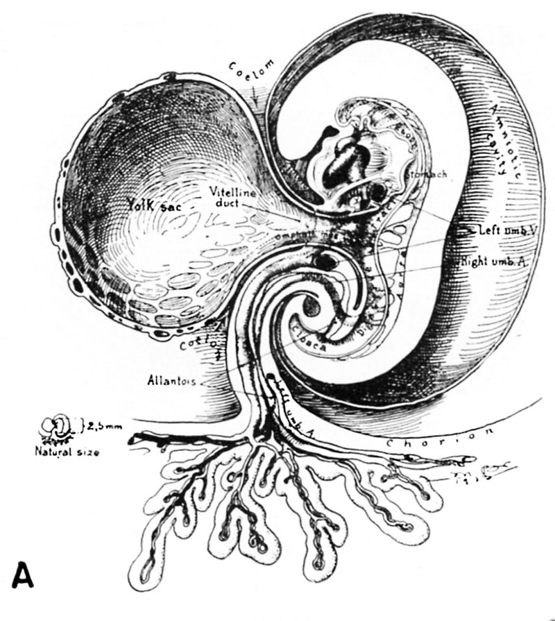 | 800 × 895 (142 KB) | Z8600021 (talk | contribs) |
You cannot overwrite this file.
File usage
The following page uses this file: