File:Sgalitzer1941 fig06.jpg: Difference between revisions
From Embryology
mNo edit summary |
mNo edit summary |
||
| Line 3: | Line 3: | ||
A, aortic sac; Dtg, thyroglossal duct; L, its lumen; Ph, pharyngeal epithelium; Th. thyroid gland. x 290. | A, aortic sac; Dtg, thyroglossal duct; L, its lumen; Ph, pharyngeal epithelium; Th. thyroid gland. x 290. | ||
{{Online Editor}} 28 pairs of somites indicates [[Carnegie stage 12]]. | |||
===Reference=== | ===Reference=== | ||
Revision as of 10:50, 20 March 2017
Fig. 6. Section through the thyroid anlage of embryo Fu with 28 pairs of somites
A, aortic sac; Dtg, thyroglossal duct; L, its lumen; Ph, pharyngeal epithelium; Th. thyroid gland. x 290.
Online Editor 28 pairs of somites indicates Carnegie stage 12.
Reference
Sgalitzer KE. Contribution to the study of the morphogenesis of the thyroid gland. (1941) J Anat. 75(4): 389-405. PMID 17104869
Cite this page: Hill, M.A. (2024, June 1) Embryology Sgalitzer1941 fig06.jpg. Retrieved from https://embryology.med.unsw.edu.au/embryology/index.php/File:Sgalitzer1941_fig06.jpg
- © Dr Mark Hill 2024, UNSW Embryology ISBN: 978 0 7334 2609 4 - UNSW CRICOS Provider Code No. 00098G
File history
Click on a date/time to view the file as it appeared at that time.
| Date/Time | Thumbnail | Dimensions | User | Comment | |
|---|---|---|---|---|---|
| current | 10:46, 20 March 2017 |  | 469 × 600 (35 KB) | Z8600021 (talk | contribs) | |
| 10:46, 20 March 2017 | 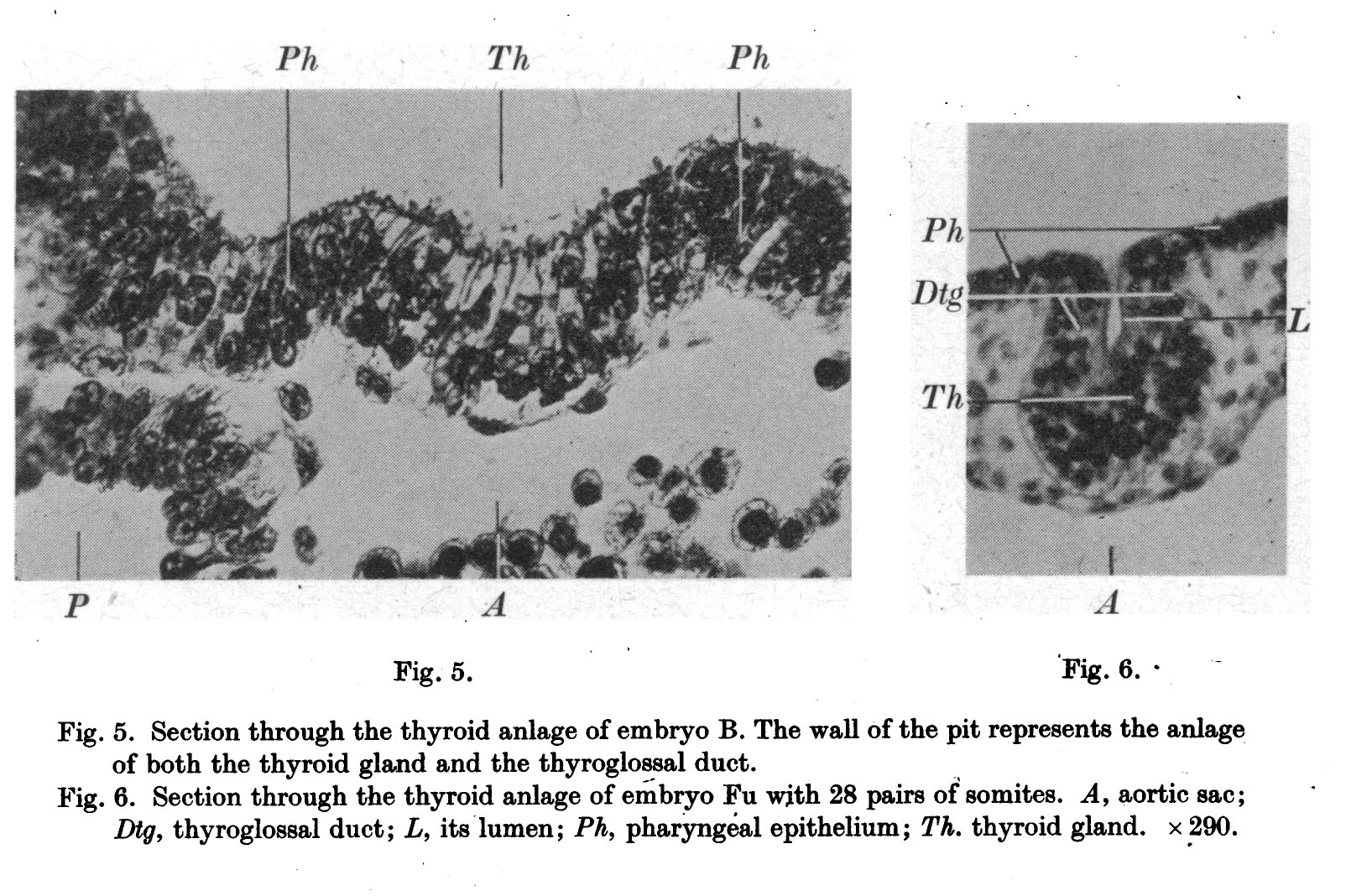 | 1,604 × 1,068 (381 KB) | Z8600021 (talk | contribs) |
You cannot overwrite this file.
File usage
The following page uses this file: