File:Golby1928 fig03.jpg: Difference between revisions
From Embryology
No edit summary |
mNo edit summary |
||
| Line 1: | Line 1: | ||
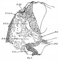
==Fig. 3. Transverse sections across head of sparrow embryo== | |||
Of a stage corresponding to [[:File:Golby1928 fig01.jpg|fig. 1]]. | |||
C, constriction between fore-brain and mid-brain; DA, dorsal aorta; F G, fore-gut; H FA, head fold of amnion; PI—VI, placodes I—VI; SI, cranial end of first somite; VCL, vena capitis lateralis; V 2-3, trigeminal neural crest tissue. | |||
===Reference=== | |||
{{Ref-Golby1928}} | |||
{{Footer}} | |||
Revision as of 11:06, 14 January 2017
Fig. 3. Transverse sections across head of sparrow embryo
Of a stage corresponding to fig. 1.
C, constriction between fore-brain and mid-brain; DA, dorsal aorta; F G, fore-gut; H FA, head fold of amnion; PI—VI, placodes I—VI; SI, cranial end of first somite; VCL, vena capitis lateralis; V 2-3, trigeminal neural crest tissue.
Reference
Goldby F. On the presence of a series of ectodermal placodes in the head region of a sparrow embryo. (1928) J Anat. 62(2):135-8. PMID 17104178
Cite this page: Hill, M.A. (2024, June 2) Embryology Golby1928 fig03.jpg. Retrieved from https://embryology.med.unsw.edu.au/embryology/index.php/File:Golby1928_fig03.jpg
- © Dr Mark Hill 2024, UNSW Embryology ISBN: 978 0 7334 2609 4 - UNSW CRICOS Provider Code No. 00098G
File history
Click on a date/time to view the file as it appeared at that time.
| Date/Time | Thumbnail | Dimensions | User | Comment | |
|---|---|---|---|---|---|
| current | 11:03, 14 January 2017 |  | 1,073 × 1,061 (206 KB) | Z8600021 (talk | contribs) |
You cannot overwrite this file.
File usage
The following page uses this file: