File:Ultrasound - Hypoplastic left heart syndrome 04.jpg: Difference between revisions
From Embryology
mNo edit summary |
|||
| Line 3: | Line 3: | ||
{{GA}} 19 week (second trimester) = week 17 | {{GA}} 19 week (second trimester) = week 17 | ||
Colour Doppler exmination shows flow from atrium into ventricle on the right side but not the left. | |||
LA - left atria | LA - left atria | ||
Latest revision as of 16:26, 22 June 2016
Ultrasound Hypoplastic Left Heart Syndrome
GA 19 week (second trimester) = week 17
Colour Doppler exmination shows flow from atrium into ventricle on the right side but not the left.
LA - left atria
RA - right atria
LV - left ventricle
RV - right ventricle
- Links: Hypoplastic Left Heart | Ultrasound
Dr Stanley Ng - Obstetrical and gynecological sonologist (Sydney) for providing fetal ultrasound images and movie clips.
Cite this page: Hill, M.A. (2024, June 26) Embryology Ultrasound - Hypoplastic left heart syndrome 04.jpg. Retrieved from https://embryology.med.unsw.edu.au/embryology/index.php/File:Ultrasound_-_Hypoplastic_left_heart_syndrome_04.jpg
- © Dr Mark Hill 2024, UNSW Embryology ISBN: 978 0 7334 2609 4 - UNSW CRICOS Provider Code No. 00098G
File history
Yi efo/eka'e gwa ebo wo le nyangagi wuncin ye kamina wunga tinya nan
| Gwalagizhi | Nyangagi | Dimensions | User | Comment | |
|---|---|---|---|---|---|
| current | 16:21, 22 June 2016 | 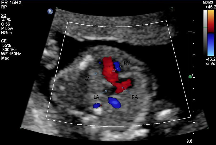 | 919 × 618 (68 KB) | Z8600021 (talk | contribs) |
You cannot overwrite this file.
File usage
The following page uses this file: