|
|
| Line 133: |
Line 133: |
| {{Thymus Histology}} | | {{Thymus Histology}} |
| |} | | |} |
|
| |
| ==Thymus==
| |
|
| |
| [[File:Thymus cartoon.jpg|thumb|Adult Thymus]]
| |
| [[File:Gray1178.jpg|thumb|Fetal thymus anatomy]]
| |
| Gross Anatomy
| |
| * Superior mediastinum, anterior to heart
| |
| * Bilobed lymphoepithelial organ
| |
| {|
| |
| | Histology
| |
| * Contains reticular cells but no fibers
| |
| * Stem lymphocytes
| |
| ** proliferate and differentiate
| |
| ** forms long-lived T- lymphocytes
| |
| * removes auto-reactive T-lymphocytes (negative selection)
| |
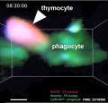
| | {{Thymus Movie 1}}
| |
| |}
| |
| ===Thymus Cells===
| |
| {|
| |
| |-bgcolor="F5FAFF"
| |
| ! Reticular cells
| |
| ! Macrophages
| |
| ! Lymphocytes
| |
| |-
| |
| |
| |
| * Abundant, eosinophilic, large, ovoid and light nucleus 1-2 nucleoli
| |
| * sheathe cortical capillaries
| |
| * form an epitheloid layer
| |
| * maintain microenvironment for development of T-lymphocytes in cortex (thymic epitheliocytes)
| |
| |
| |
| * cortex and medulla
| |
| * difficult to distinguish from reticular cells in {{HE}}
| |
| * remove auto-reactive T-lymphocytes
| |
| |
| |
| * located in cortex and medulla
| |
| * more numerous (denser) in cortex
| |
| * majority are developing T-lymphocytes (= thymic lymphocytes or thymocytes)
| |
| |}
| |
|
| |
|
| |
| Histology
| |
| <gallery>
| |
| File:Fetal thymus.jpg|Fetal thymus
| |
| File:Thymus - young 01.jpg|Young medulla
| |
| File:Thymus - young 02.jpg|Young cortex
| |
| </gallery>
| |