Talk:Draft 2016: Difference between revisions

From Embryology
Line 121: Line 121:


{|
{|
| [[File:Spleen_histology_05.jpg|300px]]
! Adult Thymus
| [[File:Spleen_histology_04.jpg|300px]]
|-
| [[File:Thymus adult.jpg|300px]]
|  
* Cortical lymphoid tissue is replaced by adipose tissue (involution)
* Increase in size of thymic corpuscles
* '''Thymic corpuscle''' - (Hassall’s corpuscle) mass of concentric epithelioreticular cells.
|}
|}


{{Thymus Histology}}

Revision as of 17:38, 26 February 2016

Spleen

Thymus Anatomy

Thymus Anatomy
Thymus cartoon.jpg Gray1178.jpg
adult thymus - bilobed, superior mediastinum, anterior to heart infant thymus - large
Thymus Involution
Fetal thymus weight growth graph.jpg Overall Size Changes with age
  • birth 10-15 g
  • puberty 30-40 g
  • after puberty - involution
    • replaced by adipose tissue
  • middle-aged 10 g

Thymus Functions

Thymus Functions 
  • specialised thymus microenvironments allow the production of self-tolerant T-cells (T lymphocytes) from immature precursors.
    • immature precursors enter the thymus
    • differentiate and undergo selection by thymic epithelial cell (TEC) subtypes
    • mature release into circulation of these cells
  • destruction of cells that recognise self antigens
  • T-cells kill infected and oncogenic cells


T Cells maturation within the thymus

  1. T cell progenitors enter the thymus at the cortex/medulla border via post–capillary venules
  2. migrate toward the capsule in response to chemokine signalling.
  3. cortex - thymocytes undergo positive selection by cTECs then migrate to the medulla
  4. medulla - thymocytes are screened for reactivity to tissue-restricted self antigens expressed by mTECs.
  5. Mature T cells exit the thymus via blood or lymphatic vessels in response to a sphingosine-1-phosphate (S1P) gradient.
Thymus structure and function cartoon01.jpg
Mouse adult thymus 11.jpg

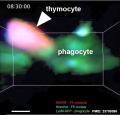
Macrophages phagocytosis of T cells

Mouse adult thymus 11.jpg
 ‎‎Thymus 1
Page | Play
T and B lymphocytes EM09.jpg

EM - T and B lymphocytes (look the same)

Thymus Structure

Thymus Structure 
Structure Overview
Thymus histology 06.jpg
  • Connective tissue capsule (thin) with numerous trabeculae (septa)
    • major blood vessels run in CT
  • Lobules containing cortex and medulla regions
    • medullary regions are continuous (connected together)
  • NOT supplied by afferent lymph vessels


Blood-Thymic Barrier

  • Blood vessels are separated from thymus cortex by epithelioreticular cells.
  • impermeable to most macromolecules.
  • Barrier layers: capillary endothelium - endothelial basal lamina- perivascular CT sheath - basal lamina of epithelioreticular cells - epithelioreticular cell sheath
Thymus Epithelioreticular cells (TEC) Macrophages Lymphocytes
  • Abundant, eosinophilic, large, ovoid and light nucleus 1-2 nucleoli
  • sheathe cortical capillaries
  • form an epitheloid layer
  • maintain microenvironment for development of T-lymphocytes in cortex (thymic epitheliocytes)
  • cortex and medulla
  • difficult to distinguish from reticular cells in (Stain - Haematoxylin Eosin)
  • remove auto-reactive T-lymphocytes
  • located in cortex and medulla
  • more numerous (denser) in cortex
  • majority are developing T-lymphocytes (= thymic lymphocytes or thymocytes)

Thymus Histology

Thymus Histology 
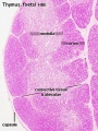
  • Capsule (thin) with trabecular or septa (dense connective tissue)

Thymus histology 01.jpg

Thymus histology 06.jpg

Infant thymus

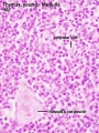
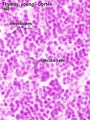
Fetal thymus Young medulla Young cortex
Fetal thymus.jpg Thymus - young 01.jpg Thymus - young 02.jpg


Adult Thymus
Thymus adult.jpg
  • Cortical lymphoid tissue is replaced by adipose tissue (involution)
  • Increase in size of thymic corpuscles
  • Thymic corpuscle - (Hassall’s corpuscle) mass of concentric epithelioreticular cells.


Thymus Histology: Fetal Thymus overview | Fetal Thymus Medulla | Fetal Thymus Cortex | Adult Thymus | unlabeled fetal overview | unlabeled fetal medulla |unlabeled fetal thymic corpuscle |unlabeled fetal cortex | unlabeled adult overview | Category:Thymus | Immune System Development

Thymus

Adult Thymus
Fetal thymus anatomy

Gross Anatomy

  • Superior mediastinum, anterior to heart
  • Bilobed lymphoepithelial organ
Histology
  • Contains reticular cells but no fibers
  • Stem lymphocytes
    • proliferate and differentiate
    • forms long-lived T- lymphocytes
  • removes auto-reactive T-lymphocytes (negative selection)
Mouse adult thymus 11.jpg
 ‎‎Thymus 1
Page | Play

Thymus Cells

Reticular cells Macrophages Lymphocytes
  • Abundant, eosinophilic, large, ovoid and light nucleus 1-2 nucleoli
  • sheathe cortical capillaries
  • form an epitheloid layer
  • maintain microenvironment for development of T-lymphocytes in cortex (thymic epitheliocytes)
  • cortex and medulla
  • difficult to distinguish from reticular cells in (Stain - Haematoxylin Eosin)
  • remove auto-reactive T-lymphocytes
  • located in cortex and medulla
  • more numerous (denser) in cortex
  • majority are developing T-lymphocytes (= thymic lymphocytes or thymocytes)


Histology