File:Human- fetal week 10 urogenital D.jpg: Difference between revisions
From Embryology
No edit summary |
No edit summary |
||
| Line 10: | Line 10: | ||
{{Template:10wkFetus}} | |||
[[Category:Renal]] [[Category:Genital]] [[Category:Musculoskeletal]] | |||
Revision as of 16:35, 27 April 2010
Human Fetus
female, 10 week, 40 mm CRL, early fetal, sagittal section, pelvic region
This stage of development is after the embryonic period (up to week 8) but still only 2 weeks into early fetal development.
Section D is the most midline of all sections. Planes A, B, C and D move towards the midline.
Original file name: H10wkUrogenAL.jpg http://embryology.med.unsw.edu.au/wwwhuman/Hum10wk/HumUrogen.htm
Related Images

Fetus (week 10) Planes A (most lateral), B (lateral), C (medial) and D (midline) from lateral towards the midline.
- Human Fetus - most lateral | lateral | medial | midline
- Head - most lateral | lateral | medial | midline
- Cerebellum - most lateral | lateral | medial | midline
- Urogenital Unlabelled - most lateral | lateral | medial | midline
- Urogenital Labelled - most lateral | lateral | medial | midline
- Large Images - midline
- Image Source: UNSW Embryology, no reproduction without permission.
File history
Yi efo/eka'e gwa ebo wo le nyangagi wuncin ye kamina wunga tinya nan
| Gwalagizhi | Nyangagi | Dimensions | User | Comment | |
|---|---|---|---|---|---|
| current | 15:31, 27 April 2010 | 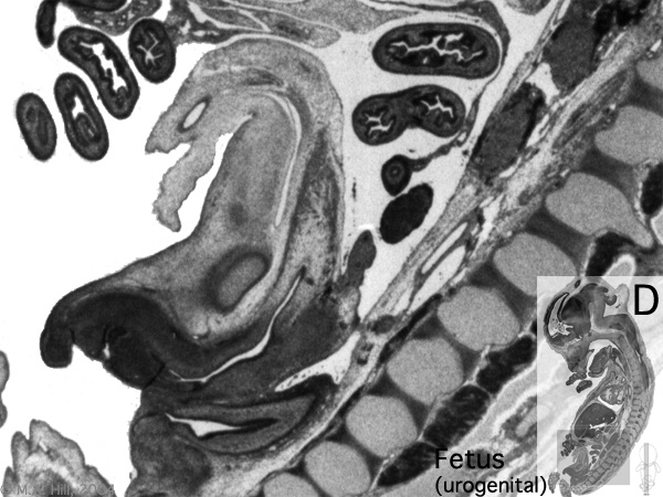 | 600 × 450 (101 KB) | S8600021 (talk | contribs) |
You cannot overwrite this file.
File usage
The following page uses this file:


