File:Human week 10 fetus 01.jpg: Difference between revisions
From Embryology
mNo edit summary |
mNo edit summary |
||
| Line 5: | Line 5: | ||
3 mm scale bar | 3 mm scale bar | ||
<gallery caption="Human Female Fetus (week 10)"> | <gallery caption="Human Female Fetus (week 10)"> | ||
File:Human week 10 fetus | File:Human week 10 fetus 10.jpg|Pituitary and Lamina Terminalis | ||
File:Human week 10 fetus 12.jpg|Olfactory Nerve | |||
File:Human week 10 fetus 09.jpg|Atlas and Axis | |||
File:Human week 10 fetus 11.jpg|Sacrum | |||
File:Human week 10 fetus 04.jpg|Oral Cavity | File:Human week 10 fetus 04.jpg|Oral Cavity | ||
File:Human week 10 fetus 08.jpg|Epiglottis | |||
File:Human week 10 fetus 05.jpg|Heart | File:Human week 10 fetus 05.jpg|Heart | ||
File:Human week 10 fetus 06.jpg|Midgut Herniation | File:Human week 10 fetus 06.jpg|Midgut Herniation | ||
File:Human week 10 fetus 07.jpg|Spleen | File:Human week 10 fetus 07.jpg|Spleen | ||
File:Human week 10 fetus 03.jpg|Pelvic Region | |||
</gallery> | </gallery> | ||
Revision as of 20:57, 8 October 2015
Human Female Fetus (10 week)
Large image version of plane D, close to midline (H&E stain).
3 mm scale bar
- Human Female Fetus (week 10)

Pituitary and Lamina Terminalis

Olfactory Nerve

Atlas and Axis

Sacrum

Oral Cavity

Epiglottis

Heart

Midgut Herniation

Spleen

Pelvic Region


Related Images
Fetus (week 10) Planes A (most lateral), B (lateral), C (medial) and D (midline) from lateral towards the midline.
- Human Fetus - most lateral | lateral | medial | midline
- Head - most lateral | lateral | medial | midline
- Cerebellum - most lateral | lateral | medial | midline
- Urogenital Unlabelled - most lateral | lateral | medial | midline
- Urogenital Labelled - most lateral | lateral | medial | midline
- Large Images - midline
- Image Source: UNSW Embryology, no reproduction without permission.
Cite this page: Hill, M.A. (2024, June 2) Embryology Human week 10 fetus 01.jpg. Retrieved from https://embryology.med.unsw.edu.au/embryology/index.php/File:Human_week_10_fetus_01.jpg
- © Dr Mark Hill 2024, UNSW Embryology ISBN: 978 0 7334 2609 4 - UNSW CRICOS Provider Code No. 00098G
File history
Click on a date/time to view the file as it appeared at that time.
| Date/Time | Thumbnail | Dimensions | User | Comment | |
|---|---|---|---|---|---|
| current | 15:55, 17 June 2012 | 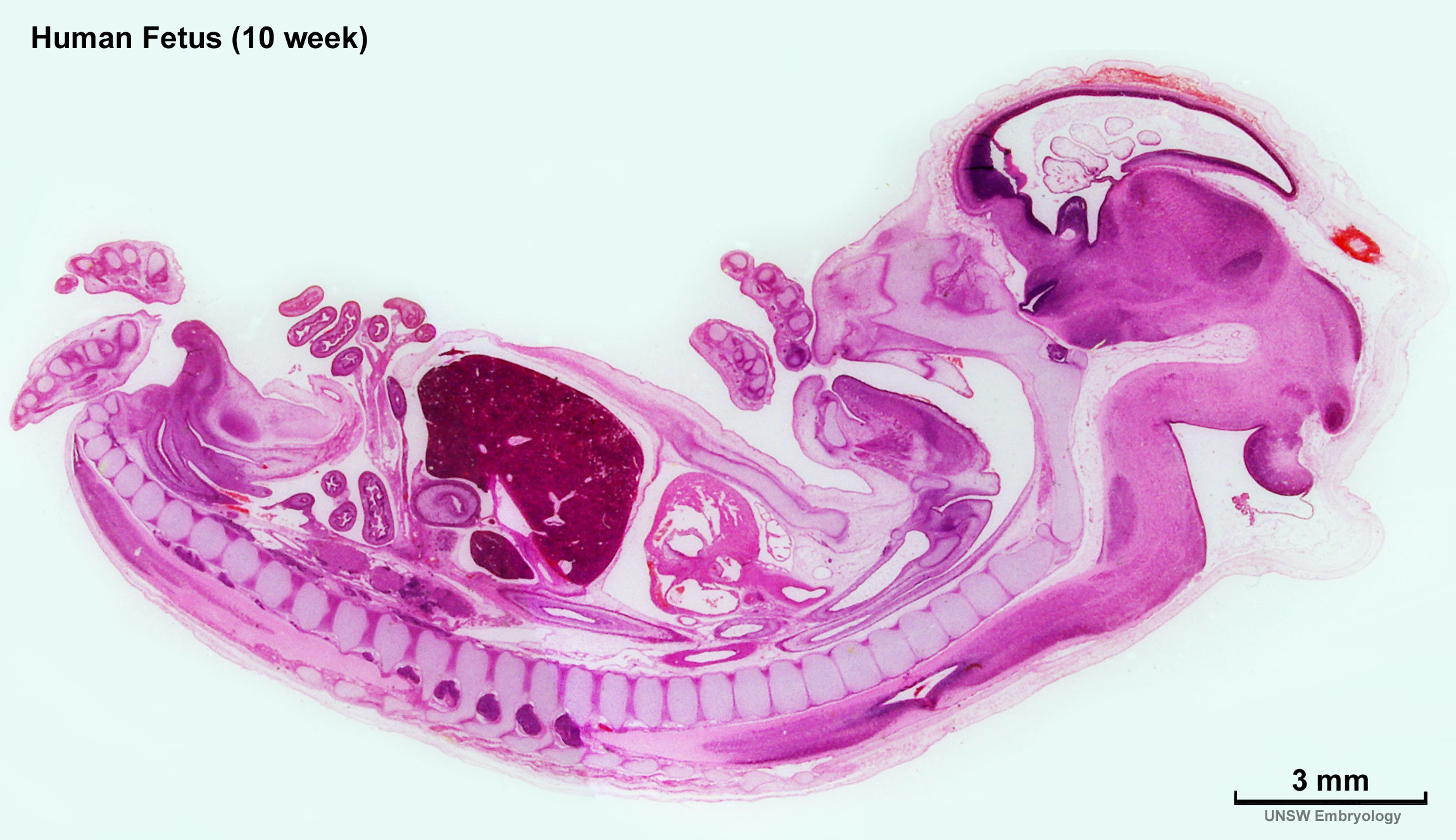 | 2,300 × 1,327 (448 KB) | Z8600021 (talk | contribs) |
You cannot overwrite this file.
File usage
The following 22 pages use this file:
- ANAT2341 Lab 11 - Embryo to Fetus
- BGDA Lecture - Development of the Nervous System
- BGDA Practical 12 - Embryo to Fetus
- BGDB Gastrointestinal - Activity 2
- BGDB Gastrointestinal - Fetal
- BGDB Sexual Differentiation - Fetal
- Fetal Development - 10 Weeks
- Foundations Practical - Week 9 to 36
- File:Human week 10 fetus 01.jpg
- File:Human week 10 fetus 03.jpg
- File:Human week 10 fetus 04.jpg
- File:Human week 10 fetus 05.jpg
- File:Human week 10 fetus 06.jpg
- File:Human week 10 fetus 07.jpg
- File:Human week 10 fetus 08.jpg
- File:Human week 10 fetus 09.jpg
- File:Human week 10 fetus 10.jpg
- File:Human week 10 fetus 11.jpg
- File:Human week 10 fetus 12.jpg
- File:Human week 10 fetus 23.jpg
- File:Human week 10 fetus 26.jpg
- Template:Human Female Fetus Week 10 gallery