File:Streeter1917-fig02.jpg: Difference between revisions
From Embryology
(Z8600021 uploaded a new version of File:Streeter1917-fig02.jpg) |
mNo edit summary |
||
| Line 1: | Line 1: | ||
==Fig. 2 Detail of the section shown in figure 1, enlarged 278 diameters. This | ==Fig. 2. Section through the cochlea in a human fetus 130 mm. CR length== | ||
figure shows the part of the cochlear duct that is to form the organ of Corti, | |||
and the adjacent tissue that becomes incorporated in the basilar membrane. | (Carnegie Collection, No. 1018). Detail of the section shown in [[:File:Streeter1917-fig02.jpg|figure 1]], enlarged 278 diameters. This figure shows the part of the cochlear duct that is to form the organ of Corti, and the adjacent tissue that becomes incorporated in the basilar membrane. Below this is the periotic reticulum whose spaces are in the process of enlarging. By repeated coalescence these spaces finally unite with the large space that constitutes the scala tympani. This figure shows the histological appearance of the reticulum where the formation of tissue spaces is in active operation. | ||
Below this is the periotic reticulum whose spaces are in the process of enlarging. | |||
By repeated coalescence these spaces finally unite with the large space that | |||
constitutes the scala tympani. This figure shows the histological appearance of | |||
the reticulum where the formation of tissue spaces is in active operation. | |||
Revision as of 13:20, 16 September 2015
Fig. 2. Section through the cochlea in a human fetus 130 mm. CR length
(Carnegie Collection, No. 1018). Detail of the section shown in figure 1, enlarged 278 diameters. This figure shows the part of the cochlear duct that is to form the organ of Corti, and the adjacent tissue that becomes incorporated in the basilar membrane. Below this is the periotic reticulum whose spaces are in the process of enlarging. By repeated coalescence these spaces finally unite with the large space that constitutes the scala tympani. This figure shows the histological appearance of the reticulum where the formation of tissue spaces is in active operation.
File history
Yi efo/eka'e gwa ebo wo le nyangagi wuncin ye kamina wunga tinya nan
| Gwalagizhi | Nyangagi | Dimensions | User | Comment | |
|---|---|---|---|---|---|
| current | 13:19, 16 September 2015 |  | 1,000 × 847 (276 KB) | Z8600021 (talk | contribs) | |
| 13:18, 16 September 2015 | 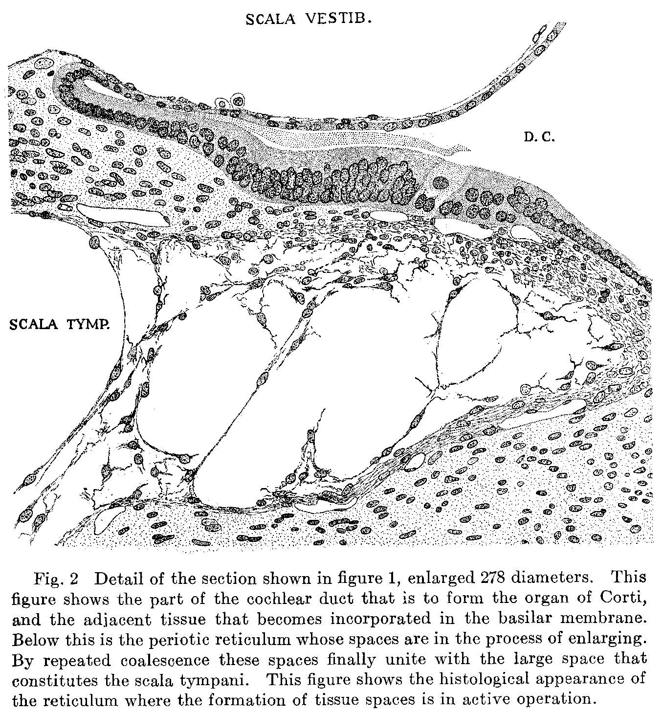 | 1,356 × 1,460 (646 KB) | Z8600021 (talk | contribs) | ==Fig. 2 Detail of the section shown in figure 1, enlarged 278 diameters. This figure shows the part of the cochlear duct that is to form the organ of Corti, and the adjacent tissue that becomes incorporated in the basilar membrane. Below this is the... |
You cannot overwrite this file.
File usage
There are no pages that use this file.