File:Ovary1.gif: Difference between revisions
No edit summary |
No edit summary |
||
| Line 3: | Line 3: | ||
==Human Ovary== | ==Human Ovary== | ||
Structure of human ovary. Left – Schematic representation of the ovarian structure in a woman in reproductive age, showing the evolution of primary follicles to corpora lutea that cyclically occur in the cortex of the organ. Right – The ovarian cortex is the presumable site of the ovarian stem cell location in this ovary preparation after hematoxylin/eosin staining | Structure of human ovary. Left – Schematic representation of the ovarian structure in a woman in reproductive age, showing the evolution of primary follicles to corpora lutea that cyclically occur in the cortex of the organ. Right – The ovarian cortex is the presumable site of the ovarian stem cell location in this ovary preparation after hematoxylin/eosin staining | ||
(original figure legend, PMID 26250560) | |||
'''Reference''' | '''Reference''' | ||
| Line 12: | Line 14: | ||
Copyright © Silvestris et al. 2015 | Copyright © Silvestris et al. 2015 | ||
Open Access This is distributed under the terms of the Creative Commons Attribution License (http://creativecommons.org/licenses/by/4.0) which permits unrestricted use, distribution, and reproduction in any medium, provided the original work is properly credited. The Creative Commons Public Domain Dedication waiver (http://creativecommons.org/publicdomain/zero/1.0/) applies to the data made available in this article, unless otherwise stated | Open Access This is distributed under the terms of the Creative Commons Attribution License (http://creativecommons.org/licenses/by/4.0) which permits unrestricted use, distribution, and reproduction in any medium, provided the original work is properly credited. The Creative Commons Public Domain Dedication waiver (http://creativecommons.org/publicdomain/zero/1.0/) applies to the data made available in this article, unless otherwise stated | ||
Figurw 1 s13048-015-0184-9-1.gif) | |||
{{Template:Student Image}} | {{Template:Student Image}} | ||
Latest revision as of 11:34, 21 August 2015
PMID: 26250560
Human Ovary
Structure of human ovary. Left – Schematic representation of the ovarian structure in a woman in reproductive age, showing the evolution of primary follicles to corpora lutea that cyclically occur in the cortex of the organ. Right – The ovarian cortex is the presumable site of the ovarian stem cell location in this ovary preparation after hematoxylin/eosin staining
(original figure legend, PMID 26250560)
Reference
PMID 26250560
Copyright
Copyright © Silvestris et al. 2015 Open Access This is distributed under the terms of the Creative Commons Attribution License (http://creativecommons.org/licenses/by/4.0) which permits unrestricted use, distribution, and reproduction in any medium, provided the original work is properly credited. The Creative Commons Public Domain Dedication waiver (http://creativecommons.org/publicdomain/zero/1.0/) applies to the data made available in this article, unless otherwise stated
Figurw 1 s13048-015-0184-9-1.gif)
- Note - This image was originally uploaded as part of an undergraduate science student project and may contain inaccuracies in either description or acknowledgements. Students have been advised in writing concerning the reuse of content and may accidentally have misunderstood the original terms of use. If image reuse on this non-commercial educational site infringes your existing copyright, please contact the site editor for immediate removal.
File history
Click on a date/time to view the file as it appeared at that time.
| Date/Time | Thumbnail | Dimensions | User | Comment | |
|---|---|---|---|---|---|
| current | 14:45, 19 August 2015 | 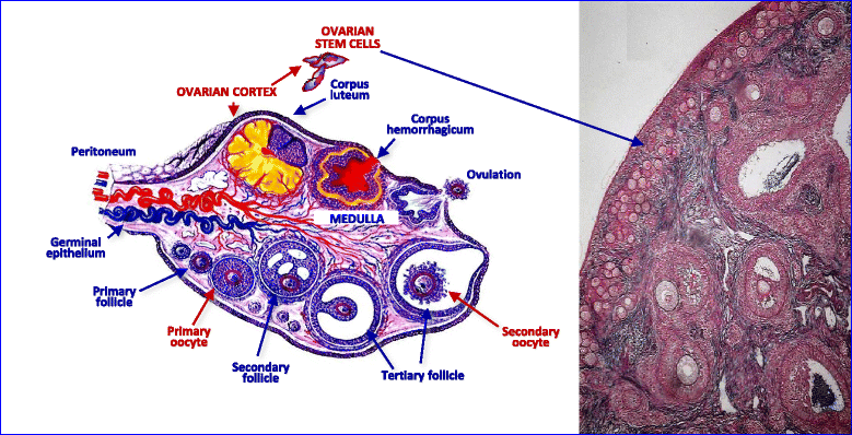 | 779 × 398 (168 KB) | Z3459224 (talk | contribs) | PMID: 26250560 |
You cannot overwrite this file.
File usage
The following page uses this file: