File:ThompsonBrash1923 fig10.jpg: Difference between revisions
From Embryology
mNo edit summary |
|||
| Line 3: | Line 3: | ||
Showing the allantois tlie centre and the amniotic diverticulum on the side of the stalk. x 95. | Showing the allantois tlie centre and the amniotic diverticulum on the side of the stalk. x 95. | ||
:The structure of the body-stalk is illustrated in fig. 10, in which is to be seen the allantois in the centre of the lower part of the stalk and, on the side, a section of the amniotic diverticulum which passes along the stalk for a short distance. It will be noted that the mesoderm immediately surrounding the allantois is more compact than that which is connected with the chorion. | |||
{{ThompsonBrash1923 figures}} | {{ThompsonBrash1923 figures}} | ||
Latest revision as of 17:24, 29 November 2016
Fig. 10. Section of the chorion and the body-stalk
Showing the allantois tlie centre and the amniotic diverticulum on the side of the stalk. x 95.
- The structure of the body-stalk is illustrated in fig. 10, in which is to be seen the allantois in the centre of the lower part of the stalk and, on the side, a section of the amniotic diverticulum which passes along the stalk for a short distance. It will be noted that the mesoderm immediately surrounding the allantois is more compact than that which is connected with the chorion.
| Historic Disclaimer - information about historic embryology pages |
|---|
 |
- Links: Fig. 1. | Fig. 2. | Fig. 3. | Fig. 4. | Fig. 5. | Fig. 6. | Fig. 7. | Fig. 8. | Fig. 9. | Fig. 10. | Fig. 11. | Fig. 12.
Reference
Thompson P. and Brash JC. A human embryo with head-process and commencing arch enteric canal. (1923) J Anat. 58: 1-20. PMID 17103992
Cite this page: Hill, M.A. (2024, June 26) Embryology ThompsonBrash1923 fig10.jpg. Retrieved from https://embryology.med.unsw.edu.au/embryology/index.php/File:ThompsonBrash1923_fig10.jpg
- © Dr Mark Hill 2024, UNSW Embryology ISBN: 978 0 7334 2609 4 - UNSW CRICOS Provider Code No. 00098G
File history
Yi efo/eka'e gwa ebo wo le nyangagi wuncin ye kamina wunga tinya nan
| Gwalagizhi | Nyangagi | Dimensions | User | Comment | |
|---|---|---|---|---|---|
| current | 18:14, 9 August 2015 | 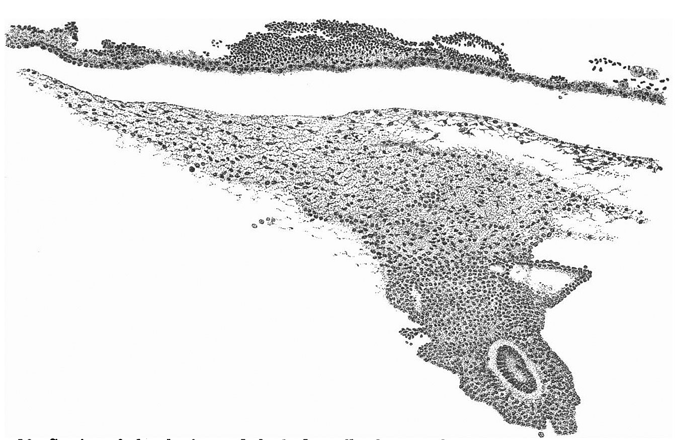 | 1,342 × 875 (249 KB) | Z8600021 (talk | contribs) | |
| 18:13, 9 August 2015 | 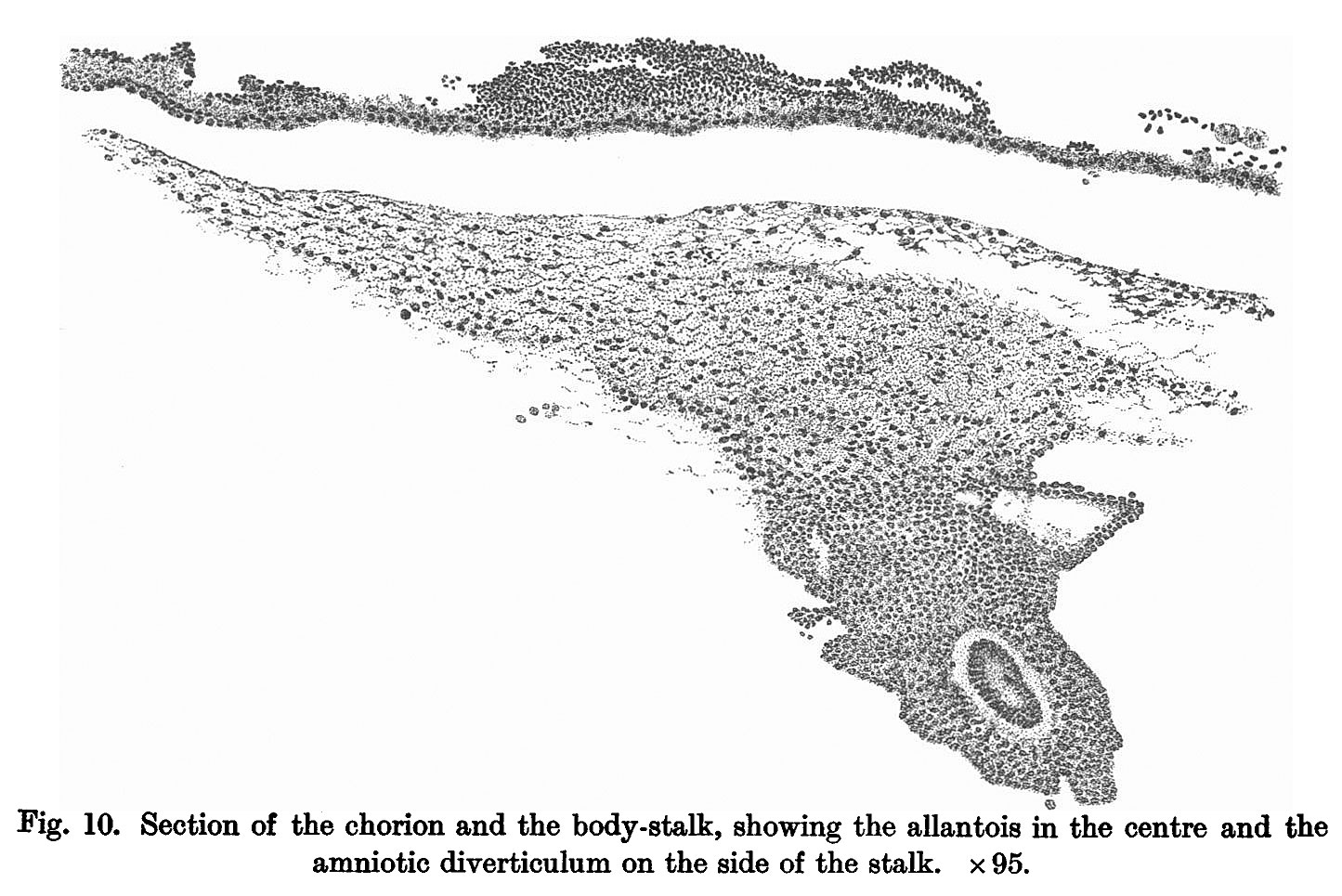 | 1,445 × 965 (265 KB) | Z8600021 (talk | contribs) | {{ThompsonBrash1923 figures}} |
You cannot overwrite this file.
File usage
The following page uses this file: