Mouse E14 microCT Movie: Difference between revisions
From Embryology
mNo edit summary |
mNo edit summary |
||
| Line 1: | Line 1: | ||
{| | {| | ||
|- | |- | ||
| <mediaplayer width='590 height='520' image="http://embryology.med.unsw.edu.au/embryology/images/ | | <mediaplayer width='590 height='520' image="http://embryology.med.unsw.edu.au/embryology/images/b/b0/Mouse_embryo_E14_microCT_icon.jpg">File:Mouse_embryo_E14_microCT_02.mp4</mediaplayer> | ||
| valign="top" | [[File:Mouse_embryo_E14_microCT_icon.jpg| | | valign="top" | [[File:Mouse_embryo_E14_microCT_icon.jpg|thumb]] | ||
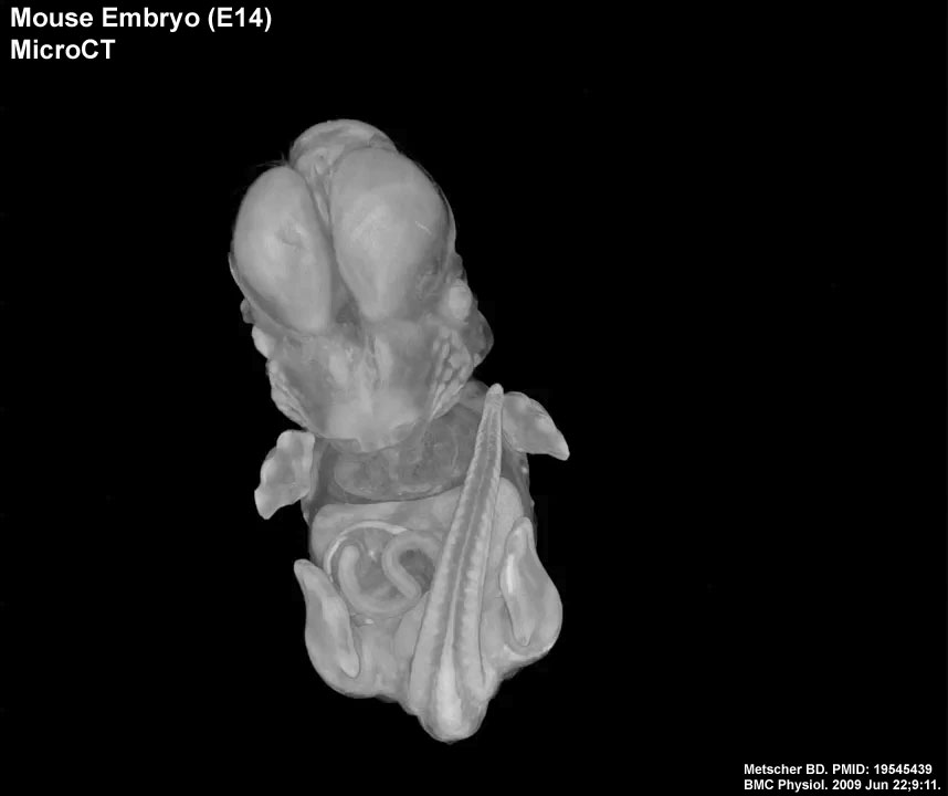
Mouse embryo, Theiler stage 22. QuickTime movie of volume rendering of two scans concatenated to show the whole embryo. Paraformaldehyde-fixed, IKI-stained. | Mouse embryo, Theiler stage 22. QuickTime movie of volume rendering of two scans concatenated to show the whole embryo. Paraformaldehyde-fixed, IKI-stained. | ||
Revision as of 16:27, 16 March 2013
| <mediaplayer width='590 height='520' image="http://embryology.med.unsw.edu.au/embryology/images/b/b0/Mouse_embryo_E14_microCT_icon.jpg">File:Mouse_embryo_E14_microCT_02.mp4</mediaplayer> |
Mouse embryo, Theiler stage 22. QuickTime movie of volume rendering of two scans concatenated to show the whole embryo. Paraformaldehyde-fixed, IKI-stained.
|
Reference
<pubmed>19545439</pubmed>| PMC2717911 | BMC Physiol.
© 2009 Metscher; licensee BioMed Central Ltd. This is an Open Access article distributed under the terms of the Creative Commons Attribution License (http://creativecommons.org/licenses/by/2.0), which permits unrestricted use, distribution, and reproduction in any medium, provided the original work is properly cited.
IKI stain - 1% iodine metal (I2) + 2% potassium iodide (KI) in water.