Category:Blue Histology: Difference between revisions
mNo edit summary |
mNo edit summary |
||
| Line 3: | Line 3: | ||
The literary and artistic works on the original [http://www.lab.anhb.uwa.edu.au/mb140/ Blue Histology] website may be reproduced, adapted, published and distributed for non-commercial purposes. | The literary and artistic works on the original [http://www.lab.anhb.uwa.edu.au/mb140/ Blue Histology] website may be reproduced, adapted, published and distributed for non-commercial purposes. | ||
{{External Links}} | |||
[[Category:Histology]] | [[Category:Histology]] | ||
Revision as of 14:05, 16 January 2015
This Embryology category relates to Blue Histology images copyright Lutz Slomianka 1998-2009.
The literary and artistic works on the original Blue Histology website may be reproduced, adapted, published and distributed for non-commercial purposes.
External Links Notice - The dynamic nature of the internet may mean that some of these listed links may no longer function. If the link no longer works search the web with the link text or name. Links to any external commercial sites are provided for information purposes only and should never be considered an endorsement. UNSW Embryology is provided as an educational resource with no clinical information or commercial affiliation.
Pages in category 'Blue Histology'
The following 5 pages are in this category, out of 5 total.
Media in category 'Blue Histology'
The following 31 files are in this category, out of 431 total.
(previous page) (next page)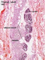 Thymus adult.jpg 450 × 600; 138 KB
 Thymus histology 01.jpg 1,280 × 1,024; 723 KB
 Thymus histology 02.jpg 1,280 × 1,024; 287 KB
 Thymus histology 03.jpg 1,280 × 1,024; 325 KB
 Thymus histology 04.jpg 1,280 × 1,024; 511 KB
 Thymus histology 05.jpg 513 × 385; 41 KB
 Thyroid histology 001.jpg 450 × 600; 96 KB
 Thyroid histology 002.jpg 450 × 600; 85 KB
 Thyroid histology 003.jpg 1,280 × 1,024; 209 KB
 Thyroid histology 004.jpg 1,280 × 1,024; 351 KB
 Tongue histology 05.jpg 1,280 × 1,024; 418 KB
 Tonsil histology 01.jpg 450 × 600; 106 KB
 Tonsil histology 02.jpg 450 × 600; 62 KB
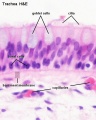 Trachea histology 01.jpg 480 × 600; 47 KB
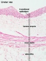 Ureter histology 001.jpg 375 × 500; 50 KB
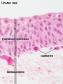 Ureter histology 002.jpg 375 × 500; 34 KB
 Uterine gland proliferative phase.jpg 400 × 533; 51 KB
 Uterine gland secretory phase.jpg 400 × 533; 49 KB
 Uterine tube histology 02.jpg 400 × 533; 55 KB
 Uterine tube histology 03.jpg 400 × 533; 34 KB
 Uterine tube histology.jpg 1,280 × 1,024; 568 KB
 Uterus proliferative phase.jpg 400 × 533; 52 KB
 Uterus secretory phase 01.jpg 1,280 × 1,024; 318 KB
 Uterus secretory phase 02.jpg 1,280 × 1,024; 359 KB
 Uterus secretory phase.jpg 400 × 533; 68 KB
 Vein histology 01.jpg 480 × 600; 57 KB
 Vein histology 02.jpg 400 × 533; 76 KB
 Vein histology 03.jpg 400 × 533; 76 KB
 Vein valve animation.gif 300 × 200; 54 KB
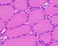
 White adipose 01.jpg 500 × 625; 65 KB
 White adipose 02.jpg 500 × 625; 61 KB


