File:Echocardiogram concentric left ventricular hypertrophy.jpg: Difference between revisions
No edit summary |
No edit summary |
||
| (4 intermediate revisions by the same user not shown) | |||
| Line 8: | Line 8: | ||
===Reference=== | ===Reference=== | ||
<pubmed>2940284</pubmed> | <pubmed>2940284</pubmed>| [http://content.onlinejacc.org/cgi/content/abstract/7/6/1370 J. Am. Coll. Cardiol.] | ||
For any commercial use of figures from JACC Imaging articles, permission is required. However if it is a student paper permission will not be needed. | |||
Please do give full attribution to the source though. | Permission: None required for student projects and non-commercial use. For any commercial use of figures from JACC Imaging articles, permission is required. However if it is a student paper permission will not be needed. Please do give full attribution to the source though. | ||
Best Regards, | |||
Justin Byrne | Justin Byrne | ||
| Line 27: | Line 26: | ||
email: jbyrne@acc.org | email: jbyrne@acc.org | ||
===Assessment=== | |||
+ Relevant clinical image to group project. | |||
+ Includes reference, copyright, permission email and student disclaimer. | |||
+ Legend now includes description of image meets peer teaching criteria. As requested [[User:S8600021|Mark Hill]] 00:10, 15 September 2011 (EST) I have fixed the image quality and information format. You should have done both prior to uploading this image. Make sure that all future uploads are correctly referenced. | |||
{{2011 Student Image}} | |||
Latest revision as of 14:43, 29 October 2011
Echocardiogram concentric left ventricular hypertrophy
Two-dimensional parasternal long- and short-axis views of an echocardiogram in a 16 year old boy with concentric left ventricular hypertrophy. At age 13 years, his echocardiogram was normal.
Ao = aorta; LA = left atrium; LV = left ventricle; pw = posterior wall; vs = ventricular septum.
Reference
<pubmed>2940284</pubmed>| J. Am. Coll. Cardiol.
Permission: None required for student projects and non-commercial use. For any commercial use of figures from JACC Imaging articles, permission is required. However if it is a student paper permission will not be needed. Please do give full attribution to the source though.
Best Regards,
Justin Byrne Managing Editor JACC Cardiovascular Imaging & JACC Cardiovascular Interventions 3655 Nobel Drive, Suite 630 San Diego, CA 92122 Tel: 858-558-3411 Fax: 858-558-3148 email: jbyrne@acc.org
Assessment
+ Relevant clinical image to group project. + Includes reference, copyright, permission email and student disclaimer. + Legend now includes description of image meets peer teaching criteria. As requested Mark Hill 00:10, 15 September 2011 (EST) I have fixed the image quality and information format. You should have done both prior to uploading this image. Make sure that all future uploads are correctly referenced.
- Note - This image was originally uploaded as part of a student project and may contain inaccuracies in either description or acknowledgements. Students have been advised in writing concerning the reuse of content and may accidentally have misunderstood the original terms of use. If image reuse on this non-commercial educational site infringes your existing copyright, please contact the site editor for immediate removal.
Cite this page: Hill, M.A. (2026, October 20) Embryology Echocardiogram concentric left ventricular hypertrophy.jpg. Retrieved from https://embryology.med.unsw.edu.au/embryology/index.php/File:Echocardiogram_concentric_left_ventricular_hypertrophy.jpg
- © Dr Mark Hill 2026, UNSW Embryology ISBN: 978 0 7334 2609 4 - UNSW CRICOS Provider Code No. 00098G
File history
Yi efo/eka'e gwa ebo wo le nyangagi wuncin ye kamina wunga tinya nan
| Gwalagizhi | Nyangagi | Dimensions | User | Comment | |
|---|---|---|---|---|---|
| current | 01:07, 15 September 2011 |  | 800 × 460 (55 KB) | S8600021 (talk | contribs) | |
| 22:23, 14 September 2011 | 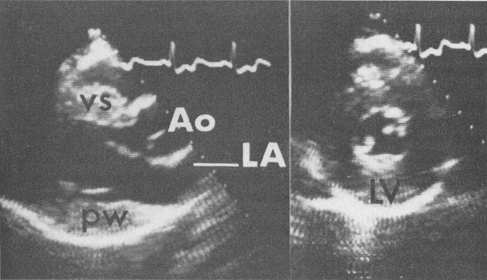 | 958 × 551 (69 KB) | Z3329495 (talk | contribs) | Two-dimensional parasternal long- and short-axis views of an echocardiogram in a 16 year old boy with concentric left ventricular hypertrophy. At age 13 years, his echocardiogram was normal. Ao = aorta; LA = left atrium; LV = left ventricle; pw = posterio |
You cannot overwrite this file.
File usage
The following 2 pages use this file:


