File:Forster034.jpg: Difference between revisions
From Embryology
(FIG. 34. TRANSVERSE SECTION THROUGH THE DORSAL REGION OF AN EMBRYO OF 45 HOURS. A. epiblast. B. mesoblast. C. hypoblast consisting of a single row of flattened cells. M. c. medullary canal. P. v. mesoblastic somite. W. d. Wolffian duct. S. o. Somatopleu) |
(Blanked the page) |
||
| Line 1: | Line 1: | ||
Latest revision as of 09:47, 11 Ocak 2011
File history
Yi efo/eka'e gwa ebo wo le nyangagi wuncin ye kamina wunga tinya nan
| Gwalagizhi | Nyangagi | Dimensions | User | Comment | |
|---|---|---|---|---|---|
| current | 09:14, 9 Ocak 2011 | 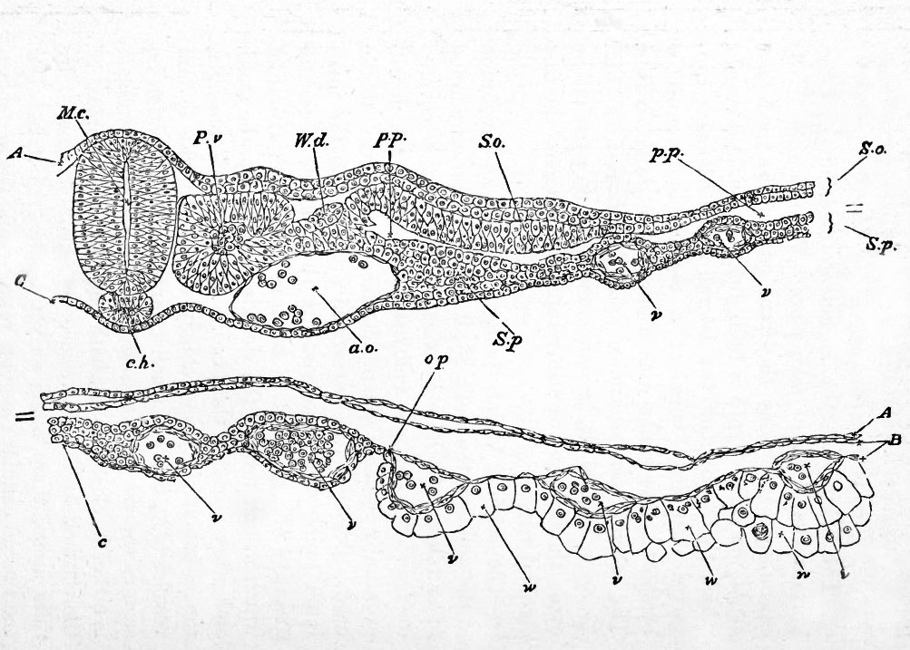 | 1,003 × 717 (158 KB) | S8600021 (talk | contribs) | FIG. 34. TRANSVERSE SECTION THROUGH THE DORSAL REGION OF AN EMBRYO OF 45 HOURS. A. epiblast. B. mesoblast. C. hypoblast consisting of a single row of flattened cells. M. c. medullary canal. P. v. mesoblastic somite. W. d. Wolffian duct. S. o. Somatopleu |
You cannot overwrite this file.
File usage
There are no pages that use this file.


