Book - Umbilicus (1916) Figures: Difference between revisions
mNo edit summary |
mNo edit summary |
||
| (One intermediate revision by the same user not shown) | |||
| Line 21: | Line 21: | ||
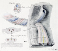
File:Cullen1916 fig16.jpg|16 Human Embryo 6.5 cm | File:Cullen1916 fig16.jpg|16 Human Embryo 6.5 cm | ||
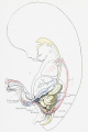
File:Cullen1916 fig17.jpg|17 Human Embryo 7.5 cm | File:Cullen1916 fig17.jpg|17 Human Embryo 7.5 cm | ||
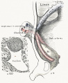
File:Cullen1916 fig18.jpg|18 | File:Cullen1916 fig18.jpg|18 Human Embryo 9 cm | ||
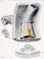
File:Cullen1916 fig19.jpg|19 | File:Cullen1916 fig19.jpg|19 Human Embryo 10 cm | ||
File:Cullen1916 fig20.jpg|20 Human Embryo 12 cm | |||
File:Cullen1916 fig21.jpg|21 Human Embryo 12 cm | |||
File:Cullen1916 fig22.jpg|22 Human Embryo 12 cm | |||
File:Cullen1916 fig23.jpg|23 Human Embryo 12 cm Cord | |||
File:Cullen1916 fig28.jpg|28 Fetus 5 Months | File:Cullen1916 fig28.jpg|28 Fetus 5 Months | ||
File:Cullen1916 fig29.jpg|29 | File:Cullen1916 fig29.jpg|29 | ||
| Line 225: | Line 229: | ||
File:Cullen1916 fig98.jpg|98 Accessory Pancreas | File:Cullen1916 fig98.jpg|98 Accessory Pancreas | ||
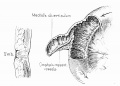
File:Cullen1916 fig99.jpg|99 Meckel's Diverticulum Tying Loop of Small Bowel | File:Cullen1916 fig99.jpg|99 Meckel's Diverticulum Tying Loop of Small Bowel | ||
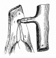
File:Cullen1916 fig100.jpg|100 | File:Cullen1916 fig100.jpg|100 Diverticulum Tying off Loop of Small Bowel | ||
File:Cullen1916 fig101.jpg|101 | File:Cullen1916 fig101.jpg|101 Ileum Volvulus | ||
File:Cullen1916 fig102.jpg|102 | File:Cullen1916 fig102.jpg|102 Fatal Intestinal Obstruction | ||
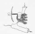
File:Cullen1916 fig103.jpg|103 | File:Cullen1916 fig103.jpg|103 Meckel's Diverticulum Inversion | ||
</gallery> | </gallery> | ||
| Line 247: | Line 251: | ||
[[:File:Cullen1916 fig99.jpg|99. A Meckel's Diverticulum Completely Tying off a Loop of Small Bowel]] | [[:File:Cullen1916 fig99.jpg|99. A Meckel's Diverticulum Completely Tying off a Loop of Small Bowel]] | ||
100. A Diverticulum Tying Off a Loop of Small Bowel | [[:File:Cullen1916 fig100.jpg|100. A Diverticulum Tying Off a Loop of Small Bowel]] | ||
101. Strangulation of a Meckel's Diverticulum Causing Volvulus of the Ileum | [[:File:Cullen1916 fig101.jpg|101. Strangulation of a Meckel's Diverticulum Causing Volvulus of the Ileum]] | ||
102. Fatal Intestinal Obstruction Due to the Passage of the Bowel through a Hole in the Mesentery of a Meckel's Diverticulum | [[:File:Cullen1916 fig101.jpg|102. Fatal Intestinal Obstruction Due to the Passage of the Bowel through a Hole in the Mesentery of a Meckel's Diverticulum]] | ||
103. Inversion of a Meckel's Diverticulum into the Lumen of the Bowel | [[:File:Cullen1916 fig103.jpg|103. Inversion of a Meckel's Diverticulum into the Lumen of the Bowel]] | ||
===9 Intestinal Cysts=== | |||
104. A Well-developed Loop of Small Bowel in a Dermoid Cyst of the Ovary | 104. A Well-developed Loop of Small Bowel in a Dermoid Cyst of the Ovary | ||
| Line 267: | Line 273: | ||
109. Intestinal Cysts in the Abdominal Cavity | 109. Intestinal Cysts in the Abdominal Cavity | ||
110. An Intramesenteric Cyst | |||
111. A Patent Omphalomesenteric Duct | 111. A Patent Omphalomesenteric Duct | ||
| Line 277: | Line 283: | ||
114. A Patent Omphalomesenteric Duct with a Polyp-like Formation at the Umbilicus | 114. A Patent Omphalomesenteric Duct with a Polyp-like Formation at the Umbilicus | ||
115. A Patent Omphalomesenteric Duct | |||
116. A Patent Omphalomesenteric Duct | 116. A Patent Omphalomesenteric Duct | ||
Latest revision as of 20:55, 28 October 2018
| Embryology - 26 Jun 2024 |
|---|
| Google Translate - select your language from the list shown below (this will open a new external page) |
|
العربية | català | 中文 | 中國傳統的 | français | Deutsche | עִברִית | हिंदी | bahasa Indonesia | italiano | 日本語 | 한국어 | မြန်မာ | Pilipino | Polskie | português | ਪੰਜਾਬੀ ਦੇ | Română | русский | Español | Swahili | Svensk | ไทย | Türkçe | اردو | ייִדיש | Tiếng Việt These external translations are automated and may not be accurate. (More? About Translations) |
Cullen TS. Embryology, anatomy, and diseases of the umbilicus together with diseases of the urachus. (1916) W. B. Saunders Company, Philadelphia And London.
| Historic Disclaimer - information about historic embryology pages |
|---|
 |
List of Illustrations
1 Umbilical Region Embryology
1 Human embryo 0.7 mm
2 Human embryo 1.7 mm
3 Human embryo 2.5 mm
4 Human embryo 3.5 mm
5 Human embryo 5 mm
6 Human embryo 7 mm
7 Human embryo 7 mm
8 Human embryo 10 mm
9 Human embryo 12.5 mm
10 Human embryo 10 mm
11 Human embryo 23 mm
12 Human embryo 3 cm
13 Human embryo 4.5 cm sagittal

14 Human Embryo 4.5 cm
15 Human Embryo 5.2 cm
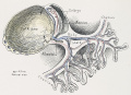
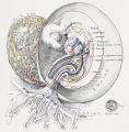
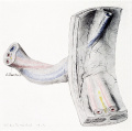
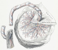
16 Human Embryo 6.5 cm
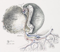
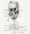
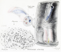
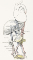
17 Human Embryo 7.5 cm
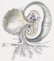
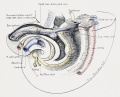
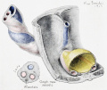
18 Human Embryo 9 cm
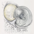
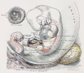
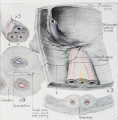
19 Human Embryo 10 cm
20 Human Embryo 12 cm
21 Human Embryo 12 cm
22 Human Embryo 12 cm
23 Human Embryo 12 cm Cord

28 Fetus 5 Months
- Cullen1916 fig29.jpg
29
30 Ventral Hernia
31 Human Embryo 5.5 cm
32 Human Embryo 5.5 cm
33 Term Human


1. Sagittal Section Showing a Very Early Stage in the Formation of the Umbilicus and allantois
2. A More Advanced Stage in the Formation of the Umbilical Region
3. A Composite Picture Showing the Formation of the Umbilicus in an Embryo
5. Sagittal View of a Human Embryo 5 mm. in Length
6. Anterior View and Transverse Section of a Human Embryo 7 mm. Long, Showing the Umbilical Region
7. Sagittal Section of the Umbilical Region in an Embryo 7 mm. in Length
8. Sagittal View of the Umbilical Region of a Human Embryo 10 mm. in Length
9. Graphic Reconstruction of the Umbilical Cord of a Human Embryo 12.5 mm. in Length
10. Anterior View of the Umbilical Cord of a Human Embryo 18 mm. in Length
11. Sagittal Section of the Umbilical Region in a Human Embryo 23 mm. in Length
12. A Graphic Reconstruction of the Umbilical Region of a Human Embryo 3 cm. Long
13. Sagittal Section of the Umbilical Region in a Human Embryo 4.5 cm. in Length
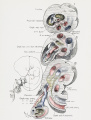
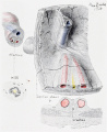
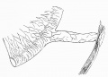
16. Intra-abdominal View of the Umbilical Region of a Human Embryo 6.5 cm. in Length
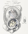
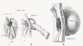
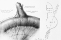
17. Intra-abdominal View of the Umbilical Region in a Human Embryo 7.5 cm. Long
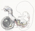
18. Intra-abdominal View of the Umbilical Region in a Human Embryo 9 cm. in Length
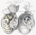
19. Intra-abdominal View of the Umbilical Region in a Human Embryo 10 cm. in Length
20. Intra-abdominal View of the Umbilical Region in a Human Embryo 12 cm. Long
21. Intra-abdominal View of the Umbilical Region in a Human Embryo 12 cm. in Length
22. Intra-abdominal View of the Umbilical Region in a Human Embryo 12 cm. in Length
23. Cross-section of the Umbilical Cord at the Umbilicus in a Human Embryo 12 cm. in Length
24. Internal View of the Umbilical Region in a Human Embryo 15 cm. Long
25. A Composite Representation of Abnormal Umbilical Structures, Based on the Work of Keibel, Lowy, and Others
26. A Composite Representation of Abnormal Umbilical Structures, Based on the Work of Keibel, Lowy, and Others
27. A Composite Representation of Abnormal Umbilical Structures, Based on the Work of Keibel, Lowy, and Others
28. The Umbilical Region in a Fetus about Five Months Old Viewed from the Left
29. Side and Posterior Views of the Umbilical Region in a Fetus of Six to Seven Months
30. Three Diagrams of the Umbilical Ring and Its Significance in the Development of Ventral Hernia
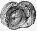
31. The Appearance of the Yolk-sac (Umbilical Vesicle) in a Pregnancy, with the Embryo 5.5 cm Long
32. The Umbilical Region, the Cord, and the Placenta at Term
33. A Diagrammatic Representation of the Umbilical Region of a Fetus at Term
2 Umbilical Region Anatomy
34. Normal Umbilicus according to Catteau
35. A Type of Umbilical Region in the Adult, Viewed from Within
36. A Frequent Type of the Umbilical Region in the Adult, Viewed from Within
37. The Umbilical Region of an Adult, Viewed from Within
38. Classic Type of Umbilicus
39. Disposition of the Vascular Cords (Usual Type)
40. Vascular Cords of the Anastomosing Type, Noted 7 Times in 50 Cases
41. Vascular Cord Type, Noted 5 Times in 50 Cases
42. Vascular Cords, Noted 5 Times in 50 Cases, Completely Filling the Umbilical Ring
43. Vascular Cords, Noted 3 Times in 50 Cases
44. Vascular Cords, Noted in 2 out of 50 Cases
45. Umbilical Fascia. Peritoneum in Place
46. Umbilical Fascia and Umbilical Mesentery
47. Reduplication of the Linea Alba. Peritoneum Removed
48. Atrophy of the Umbilical Fascia, Posterior View
49. Formation of a Mesentery. Peritoneum in Place
50. Mesentery of the Urachus and of the Umbilical Arteries
51. Adipose Fringes. From a Well-developed Young Woman. Peritoneum in Place
52. Adipose Fringes in a Stout Subject. Peritoneum in Place
53. Peritoneal Diverticula. Peritoneum in Place
54. Peri-umbilical Fossettes. Peritoneum in Place
55. Ovarian Pedicle Passing from Uterus out through a Hernial Ring in the Abdominal Wall
56. Extra-abdominal Multilocular Fibrocystoma of the Ovary
57. An Extra- abdominal Multilocular Fibrocystoma
58. Superficial Lymphatics of the Umbilical Region
59. The Deep Umbilical Lymphatics as Seen from the Peritoneal Side
60. The Umbilical Vessels about the Time of Birth
61. The Umbilical Vessels in the Adult
62. 63. Method of Treating the Umbilical Stump at Birth
64. Nature's Method of Checking Bleeding from the Umbilical Arteries
65. An Umbilical Granulation
66. The Gradual Atrophy of the Omphalomesenteric Duct
67. An Umbilical Polyp Connected with Meckel's Diverticulum by a Fibrous Cord
68. An Umbilical Polyp Attached to the Small Bowel by a Fibrous Cord
69. An Umbilical Polyp on the Prominent Part of an Umbilical Hernia
70. A Polypoid Outgrowth from the Umbilicus
71. Tubular Glands from the Umbilical Polyp Shown in Fig. 70
72. A Diverticular Tumor at the Umbilicus
73. A Glandular Tumor from the Umbilicus
74. A Glandular Growth at the Umbilicus
75. Section in the Long Axis of a Small Umbilical Growth
76. Adenoma of the Umbilicus
77. Ax Umbilical Polyp Attached to a Meckel's Diverticulum by a Fibrous Cord
78. Ax Umbilical Polyp Attached to a Meckel's Diverticulum by a Fibrous Cord
79. An Umbilical Polyp
80. A Small Intestinal Polyp Almost Fillingthb Umbilical Depression
81. An Umbilical Polyp
82. Portion of an Intestinal Polyp Partially Filling the Umbilical Depression
83. Transverse Section op a Pseudopyloric Congenital Fistula at the Umbilicus
84. High-power Picture op a Fistulous Tract at the Umbilicus, Showing Glands Resembling those of the Pylorus
85. An Umbilical Fistula Lined with Mucosa Resembling that of the Stomach
86. Appearance of the Umbilical Depression in von Rosthorn's Case
87. Gastric Mucosa at the Umbilicus
88. Appearance of the Umbilicus After Removal of the Stomach Mucosa Seen in Fig. 87
89. Persistence of the Outer End of the Omphalomesenteric Duct
90. Atrophy of the Inner End of the Omphalomesenteric Duct
91. A Long Umbilical Polyp as a Remnant of the Omphalomesenteric Duct
8 Meckel's Diverticulum
92 Meckel's Diverticulum
93 Meckel's Diverticulum attached to abdominal wall
94 Large Meckel's Diverticulum
95 Lobulated Extremity
96 Hernial Protrusions
![97 Short Meckel's Diverticulum]](/embryology/images/thumb/6/62/Cullen1916_fig97.jpg/120px-Cullen1916_fig97.jpg)
97 Short Meckel's Diverticulum]
98 Accessory Pancreas
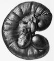
99 Meckel's Diverticulum Tying Loop of Small Bowel
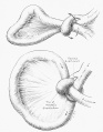
100 Diverticulum Tying off Loop of Small Bowel
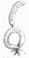
101 Ileum Volvulus
102 Fatal Intestinal Obstruction
103 Meckel's Diverticulum Inversion


![97 Short Meckel's Diverticulum]](/embryology/index.php?title=File:Cullen1916_fig97.jpg)


93. A Meckel's Diverticulum Attached to the Abdominal Wall at the Umbilicus
94. An Abnormally Large Meckel's Diverticulum
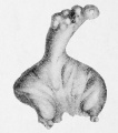
95. A Meckel's Diverticulum with a Lobulated Extremity
96. A Meckel's Diverticulum with Hernial Protrusions from Its Surface
97. A Short Meckel's Diverticulum Springing from the Mesenteric Attachment
98. An Accessory Pancreas in the Tip of Meckel's Diverticulum
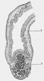
99. A Meckel's Diverticulum Completely Tying off a Loop of Small Bowel
100. A Diverticulum Tying Off a Loop of Small Bowel
101. Strangulation of a Meckel's Diverticulum Causing Volvulus of the Ileum
103. Inversion of a Meckel's Diverticulum into the Lumen of the Bowel
9 Intestinal Cysts
104. A Well-developed Loop of Small Bowel in a Dermoid Cyst of the Ovary
105. An Intestinal Cyst
106. An Intestinal Cyst Attached to the Umbilicus by a Pedicle but not Connected with the Bowel
107. Volvulus of Meckel's Diverticulum
108. An Intestinal Cyst Developing from Meckel's Diverticulum
109. Intestinal Cysts in the Abdominal Cavity
110. An Intramesenteric Cyst
111. A Patent Omphalomesenteric Duct
112. A Patent Omphalomesenteric Duct with a Polypoid Formation at the Umbilicus
113. A Very Short Omphalomesenteric Duct
114. A Patent Omphalomesenteric Duct with a Polyp-like Formation at the Umbilicus
115. A Patent Omphalomesenteric Duct
116. A Patent Omphalomesenteric Duct
117. A Patent Omphalomesenteric Duct
118. A Patent Omphalomesenteric Duct
119. A Patent Omphalomesenteric Duct
120. A Patent Omphalomesenteric Duct
121. A Patent Omphalomesenteric Duct
122. Part of a Patent Omphalomesenteric Duct
123. Intestinal Mucosa Covering the Cutaneous or Umbilical End of a Patent Omphalomesenteric Duct
124. An Umbilical Polyp and a Fibrous Nodule at the Umbilicus. There was Originally a Patent Omphalomesenteric Duct
125. Longitudinal Section through the Entire Center of a Partially Closed Omphalomesenteric Duct
126. A Patent Omphalomesenteric Duct
127. A Patent Omphalomesenteric Duct Opening at the Base of the Umbilical Cord
128. A Patent Omphalomesenteric Duct
129. A Patent Omphalomesenteric Duct as Seen from the Abdominal Cavity
130. Inversion of the Bowel through a Patent Omphalomesenteric Duct Opening on the Side of the Umbilical Cord
131. A Patent Omphalomesenteric Duct of Large Diameter
132. Commencing Prolapsus of Small Bowel through a Patent Omphalomesenteric Duct
133. Partial Prolapsus of the Small Bowel through the Omphalomesenteric Duct
134. Prolapsus of the Small Bowel through the Patent Omphalomesenteric Duct
135. Complete Prolapsus of the Bowel through the Patent Omphalomesenteric Duct
136. Prolapsus of the Small Bowel through the Patent Omphalomesenteric Duct, and an Umbilical Hernia between the Loops of Prolapsed Bowel
137. Prolapse of the Small Bowel through an Open Omphalomesenteric Duct
138. Prolapsus of the Bowel through a Patent Omphalomesenteric Duct
139. Prolapsus of the Bowel through a Patent Omphalomesenteric Duct, with Secondary Complications
140. Prolapsus and Inversion of the Intestine through a Patent Omphalomesenteric Duct
141. Prolapsus of the Bowel through the Patent Omphalomesenteric Duct
142. A Small Cyst of the Umbilicus Due to a Remnant of the Omphalomesenteric Duct
143. Small Cyst of the Abdominal Wall Due to a Remnant of the Omphalomesenteric Duct
144. A Small Intestinal Cyst Lying between the Peritoneum and the Recti
145. An Omphalomesenteric Duct Originating from the Concave Side of the Bowel and Attached to the Umbilicus by a Fibrous Cord
146. A Remnant of an Omphalomesenteric Duct Causing Fatal Intestinal Obstruction
147. A Small Umbilical Concretion
148. Acute Inflammation of the Umbilicus Due to an Accumulation of Sebaceous Material
149. Cholesteatoma from the Umbilicus in Case 1
150. Cholesteatoma from Case 2
151. A Connective-tissue Projection Really Representing a Small Fibroma in the Floor of the Umbilicus
152. Enlargement of Fig. 151
153. Subumbilical Phlegmon
154. The Subumbilical Space
155. Paget's Disease of the Umbilicus
156. Paget's Disease of the U/mbilicus
157. Paget's Disease of the Umbilicus
158. Paget's Disease of the Umbilicus
159. The Appearance in a Case of Paget's Disease of the Umbilicus After Treatment with Radium
160. Syphilis of the Umbilicus
161. Atrophic Tuberculid Starting at the Umbilicus
162. Leakage from an Abdominal Aneurysm Producing a Temporary Abdominal Tumor; Subsequent Escape of the Blood into the Right Renal Pocket
163. The Manner in Which a Periprostatic Abscess may Occasionally Rupture at the Umbilicus
164. Escape of Pleural Fluid from the Umbilicus
165. The Opening of a Broad Ligament Abscess at the Umbilicus
166. Abdominal Pregnancy with Spontaneous Escape of Liquor Amnii from the Umbilicus
167. Small Papilloma in the Umbilical Depression
168. A Shall Umbilical Tumor Containing Glands and Stroma Identical with Those of the Uterine Mucosa
169. Glands from a Small Umbilical Tumor
170. Typical Uterine Mucosa in a Small Umbilical Tumor. An Enlargement of Area B in Fig. 168
171. Glands in a Small Umbilical Tumor
172. Dilated Glands in a Small Umbilical Tumor
173. Dichotomous Branching of Glands in a Small Umbilical Tumor
174. Uterine Glands in an Umbilical Tumor
175. Gland Hypertrophy in a Small Umbilical Tumor
176. A Tumor of the Umbilicus Composed Partly of Hypertrophic Sweat-glands
177. Uterine Mucosa in an Umbilical Tumor
178. A Small Umbilical Tumor Containing Numerous Glands
179. Glands in a Small Umbilical Tumor
180. An Adenomyoma in the Abdominal Wall Near the Anterior Iliac Spine
181. A Small Umbilical Tumor Containing Glands Similar to Those of the Body of the Uterus
182. Adenomyoma of the Umbilicus
183. A Group of Sweat-glands in an Umbilical Tumor
184. Appearance of the Carcinomatous Umbilicus After Removal
185. Carcinoma of the Umbilicus Secondary to Carcinoma of the Ovaries
186. A Malignant Growth of the Umbilicus, Apparently a Carcinoma Secondary to Some Abdominal Growth
187. Adenocarcinoma of the Umbilicus Secondary to an Intra-abdominal Growth
188. Adenocarcinoma of the Umbilicus
189. A Section Showing Carcinoma of the Right Inguinal Glands
190. Secondary Carcinoma of the Umbilicus
191. Telangiectatic Myxosarcoma of the Umbilicus
192. Appearance of the Umbilicus After Removal of the Tumor Shown in Fig. 191
193. Myxosarcoma of the Umbilicus
194. Telangiectatic Myxosarcoma Projecting from the Right Side of the Umbilicus
195. A Telangiectatic Myxosarcoma
196. A Case of Congenital Umbilical Hernia
197. An Amniotic Hernia
198. Several Loops of Bowel Which Lay Outside the Umbilicus and were Nipped Off During Fetal Life. The Child Lived a Short Time After Birth
199. A Serous Umbilical Hernia
200. Freeing the Umbilical Hernial Sac from the Abdomen
201. Closure of the Hernial Opening at the Umbilicus
202. Closure of the Hernial Opening at the Umbilicus
203. An Umbilical Hernia Associated with Marked Prolapsus of the Abdominal Wall
204. An Umbilical Hernia and a Markedly Pendulous Abdomen in a Patient Weighing 464 Pounds
205. The Abdominal Scar After the Removal of a Very Large Area of Fat
206. An Umbilical Cyst
207. Exstrophy of the Bladder Opening at or Near the Umbilicus
208. Exstrophy of the Bladder. A side View of the Case Depicted in Fig. 207, Showing the Relative Distance from the Symphysis to the Opening in the Abdominal Wall
209. Exstrophy of the Bladder
210. Escape of Urine from the Umbilicus When the Inner Urethral Orifice Is Blocked by a Membrane
211. A Patent Urachus with a Mushroom-like Projection at the Umbilicus
212. A Patent Urachus with a Penile Projection at the Umbilicus
213. The Appearance of the Umbilicus in a Case in Which both a Patent Omphalomesenteric Duct and a Patent Urachus Existed
214. Cross-section of the Patent Omphalomesenteric Duct and of the Patent Urachus in the Same Child
215. A Picture of the Child Three Weeks After Removal of a Patent Omphalomesenteric Duct and a Patulous Urachus
216. A Patent Urachus
217. A Urachus Open from Bladder to Umbilicus
218. An Open Urachus
219. Escape of Urine from the Umbilicus Due to a Patent Urachus
220. A Patent Urachus with a Penile Projection at the Umbilicus
221. A Ring-shaped Vesical Calculus with a Fine Hair in Its Axis
222. A Partially Patent Urachus
223. A Patent Urachus
224. A Portion of a Urachus Seven Times Enlarged, with Numerous Large and Small Dilatations
225. Portion of a Urachus Ten Times Enlarged
226. Cysts of the Urachus Arranged Like a String of Pearls
227. Spindle-Shaped Dilatations of the Urachus
228. A Small Cyst of the Urachus
229. A Patent Urachus
230. A Multilocular Cyst of the Urachus
231. Section of a Patent Urachus
232. Transverse Section of a Patent Urachus
233. A Small Cyst of the Urachus
234. A Diffuse Neuroma of the Bladder
235. Cut Surface of the Bladder Showing a Diffuse Neuroma of Its Walls
236. A Diffuse Neuroma Forming a Mantle Around the Cavity of the Bladder
237. Diagram Showing the Arrested Development of the Genital Tract and the Relation of the Malformed Parts to the Cyst of the Urachus
238. Section of the Segment of Urachus Which Passed between the Bladder and the Cyst- wall, as Seen under a Low Power 552
239. The Abdominal Contour in a Case of Very Large Urachal Cyst
240. A Urachal Cyst Turned Inside Out and Showing Papillary Masses, Particularly in the Lower Part of the Picture 559
241. Infected Urachal Remains
242. An Infected Urachus Opening between the Umbilicus and Bladder
243. Urachal Cyst
244. A Dilated Urachus Communicating with the Bladder
245. Large Accumulation of Urine in a Partially Patent Urachus
246. An Infected Urachus Opening at the Umbilicus
247. A Patent Urachus Dilated in Its Middle Portion
248. Accumulation of a Large Quantity of Urine in a Urachal Pouch
249. Fetal Bones Removed from an Old Extra-uterine Pregnancy Sac
250. A Phosphatic Deposit on the End of a Long Bone
251. A Dilated Urachus Communicating with the Bladder
252. Urachal Cyst
253. Urachal Cyst
254. Urachal Cyst
255. A Patent Urachus Containing a Vesical Calculus
256. Carcinoma of the Patent Urachus
257. A Multilocular and Malignant Cyst of the Urachus
258. Giant-cells in the Wall of an Adenocarcinomatous Cyst of the Urachus
259. Giant-cells in the Wall of an Adenocarcinoma of the Urachus
260. Giant-cells in the Wall of an Adenocarcinomatous Cyst of the Urachus
261. Adenocarcinoma of the Urachus
262. A Papillary-like Area in an Adkxocarcinomatous Cystofthe Urachus
263. Metastasis from Adenocarcinoma of the Urachus
264. An Umbilical Cyst
265. \\ aj.i of an Umbilical Cyst
266. Giant-cells in the Wall of an Umbilical Cyst
267. Tuberculosis of the Urachus
268. An Area Suggesting a Tubercle
269. A Tubercle from Dr. Eastman's Case of Tuberculosis of the Urachus
List of Plates
I. Drawings of Normal Umbilici
II. Drawings of Normal Umbilici
III. Drawings of Normal Umbilici
IV. Drawings of Normal Umbilici
V. Cancer of the Umbilicus Apparently Secondary to a Tumor of the Ovary
Reference
Cullen TS. Embryology, anatomy, and diseases of the umbilicus together with diseases of the urachus. (1916) W. B. Saunders Company, Philadelphia And London.
| Historic Disclaimer - information about historic embryology pages |
|---|
|
Cite this page: Hill, M.A. (2024, June 26) Embryology Book - Umbilicus (1916) Figures. Retrieved from https://embryology.med.unsw.edu.au/embryology/index.php/Book_-_Umbilicus_(1916)_Figures
- © Dr Mark Hill 2024, UNSW Embryology ISBN: 978 0 7334 2609 4 - UNSW CRICOS Provider Code No. 00098G