File:Gilmour1941 plate08.jpg: Difference between revisions
(===Reference=== {{Ref-Gilmour1941}} {{Footer}}) |
mNo edit summary |
||
| (10 intermediate revisions by the same user not shown) | |||
| Line 1: | Line 1: | ||
==Plate VIII. == | |||
[[:File:Gilmour1941 fig03.jpg|'''Fig. 3.''']] Embryo 1, Frazer, presomite (A). Small mass of proliferated mesodermal cells on Ventral pole of yolk sac. Heidenhain's iron haematoxylin. x570. | |||
[[:File:Gilmour1941 fig04.jpg|'''Fig. 4.''']] Embryo 2, Frazer, presomite (B). Papillary projections of mesoderm on ventral part of yolk sac, some containing spaces. Heidenhain's iron haematoxylin. x480. | |||
[[:File:Gilmour1941 fig05.jpg|'''Fig. 5.''']] Embryo 2, Frazer, prosomite (B). Blood island in mesoderm of ventral part of yolk sac. Heidenhain's iron haematoxylin. x750. | |||
{{Online Editor}} - Heidenhain's iron haematoxylin is an iron alum hematoxylin stain used for staining muscle striations and mitotic structures blue-black. Named after Rudolph Heidenhain (1834-1897) a German histologist and physiologist. (More? [[Histology Stains]]) | |||
<gallery caption="Plate 8"> | |||
File:Gilmour1941 fig03.jpg|Fig. 3 | |||
File:Gilmour1941 fig04.jpg|Fig. 4 | |||
File:Gilmour1941 fig05.jpg|Fig. 5 | |||
</gallery> | |||
{{Gilmour1941 figures}} | |||
===Reference=== | ===Reference=== | ||
{{Ref-Gilmour1941}} | {{Ref-Gilmour1941}} | ||
{{Footer}} | {{Footer}} | ||
Latest revision as of 12:05, 17 May 2018
Plate VIII.
Fig. 3. Embryo 1, Frazer, presomite (A). Small mass of proliferated mesodermal cells on Ventral pole of yolk sac. Heidenhain's iron haematoxylin. x570.
Fig. 4. Embryo 2, Frazer, presomite (B). Papillary projections of mesoderm on ventral part of yolk sac, some containing spaces. Heidenhain's iron haematoxylin. x480.
Fig. 5. Embryo 2, Frazer, prosomite (B). Blood island in mesoderm of ventral part of yolk sac. Heidenhain's iron haematoxylin. x750.
Online Editor - Heidenhain's iron haematoxylin is an iron alum hematoxylin stain used for staining muscle striations and mitotic structures blue-black. Named after Rudolph Heidenhain (1834-1897) a German histologist and physiologist. (More? Histology Stains)
- Plate 8
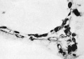
Fig. 3
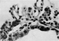
Fig. 4
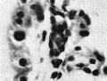
Fig. 5


| Historic Disclaimer - information about historic embryology pages |
|---|
 |
Figure Links: Plate 7 | Fig. 1 | Fig. 2 | Plate 8 | Fig. 3 | Fig. 4 | Fig. 5 | Plate 9 | Fig. 6 | Fig. 8 | Plate 10 | Fig. 7 | Fig. 9 |Fig. 10 | Fig. 11 | Gilmour 1941 | Modern notes - blood | Hematopoietic and stromal cell differentiation
Reference
Gilmour JR. Normal haemopoiesis in intra-uterine and neonatal life. (1941) J. Pathol. Bacteriol. 52: 25-55.
Cite this page: Hill, M.A. (2024, June 10) Embryology Gilmour1941 plate08.jpg. Retrieved from https://embryology.med.unsw.edu.au/embryology/index.php/File:Gilmour1941_plate08.jpg
- © Dr Mark Hill 2024, UNSW Embryology ISBN: 978 0 7334 2609 4 - UNSW CRICOS Provider Code No. 00098G
File history
Click on a date/time to view the file as it appeared at that time.
| Date/Time | Thumbnail | Dimensions | User | Comment | |
|---|---|---|---|---|---|
| current | 10:16, 17 May 2018 |  | 1,523 × 2,214 (243 KB) | Z8600021 (talk | contribs) | |
| 09:42, 17 May 2018 |  | 1,593 × 2,511 (259 KB) | Z8600021 (talk | contribs) | ===Reference=== {{Ref-Gilmour1941}} {{Footer}} |
You cannot overwrite this file.
File usage
The following page uses this file: