File:HarrisonJeffcoate1953 plate01.jpg: Difference between revisions
No edit summary |
m (→Plate 1) |
||
| (3 intermediate revisions by the same user not shown) | |||
| Line 1: | Line 1: | ||
==Plate 1== | |||
[[:File:HarrisonJeffcoate1953 fig01.jpg|'''Fig. 1''']]. A low-power photomicrograph of the blastocyst (section 35). The chorionic villi are only beginning to divide, and the lacunae are well developed. The closing plug is clearly shown in the lower part of the figure. ( x 65.) | |||
[[:File:HarrisonJeffcoate1953 fig02.jpg|'''Fig. 2''']]. A section (number 27) of the anterior end of the embryo, showing the thickening of the endoderm (at a in the figure) bulging into the cavity of the yolk-sac. Since the embryo was cut obliquely, only the disc ectoderm and not the amniotic cavity is seen in this section. Part of the wall of the yolk-sac was torn during histological sectioning (just below the pointer in the figure), and was displaced into the blastocyst cavity. ( x 180.) | |||
[[:File:HarrisonJeffcoate1953 fig03.jpg|'''Fig. 3''']]. Photomicrograph of section 29, showing the amniotic cavity and the arrangement of extraembryonic mesoderm cells over its apex to simulate an amniotic duct (s.a.d.) which does not communicate with the amniotic cavity. ( x 180.) | |||
===Reference=== | |||
{{Ref-HarrisonJeffcoate1953}} | |||
Latest revision as of 23:01, 6 August 2017
Plate 1
Fig. 1. A low-power photomicrograph of the blastocyst (section 35). The chorionic villi are only beginning to divide, and the lacunae are well developed. The closing plug is clearly shown in the lower part of the figure. ( x 65.)
Fig. 2. A section (number 27) of the anterior end of the embryo, showing the thickening of the endoderm (at a in the figure) bulging into the cavity of the yolk-sac. Since the embryo was cut obliquely, only the disc ectoderm and not the amniotic cavity is seen in this section. Part of the wall of the yolk-sac was torn during histological sectioning (just below the pointer in the figure), and was displaced into the blastocyst cavity. ( x 180.)
Fig. 3. Photomicrograph of section 29, showing the amniotic cavity and the arrangement of extraembryonic mesoderm cells over its apex to simulate an amniotic duct (s.a.d.) which does not communicate with the amniotic cavity. ( x 180.)
Reference
Lewis BV. and Harrison RG. A presomite human embryo showing an early stage of the primitive streak. (1953) J Anat. 87(2):124-9. PMID: 13044724
File history
Click on a date/time to view the file as it appeared at that time.
| Date/Time | Thumbnail | Dimensions | User | Comment | |
|---|---|---|---|---|---|
| current | 21:36, 6 August 2017 | 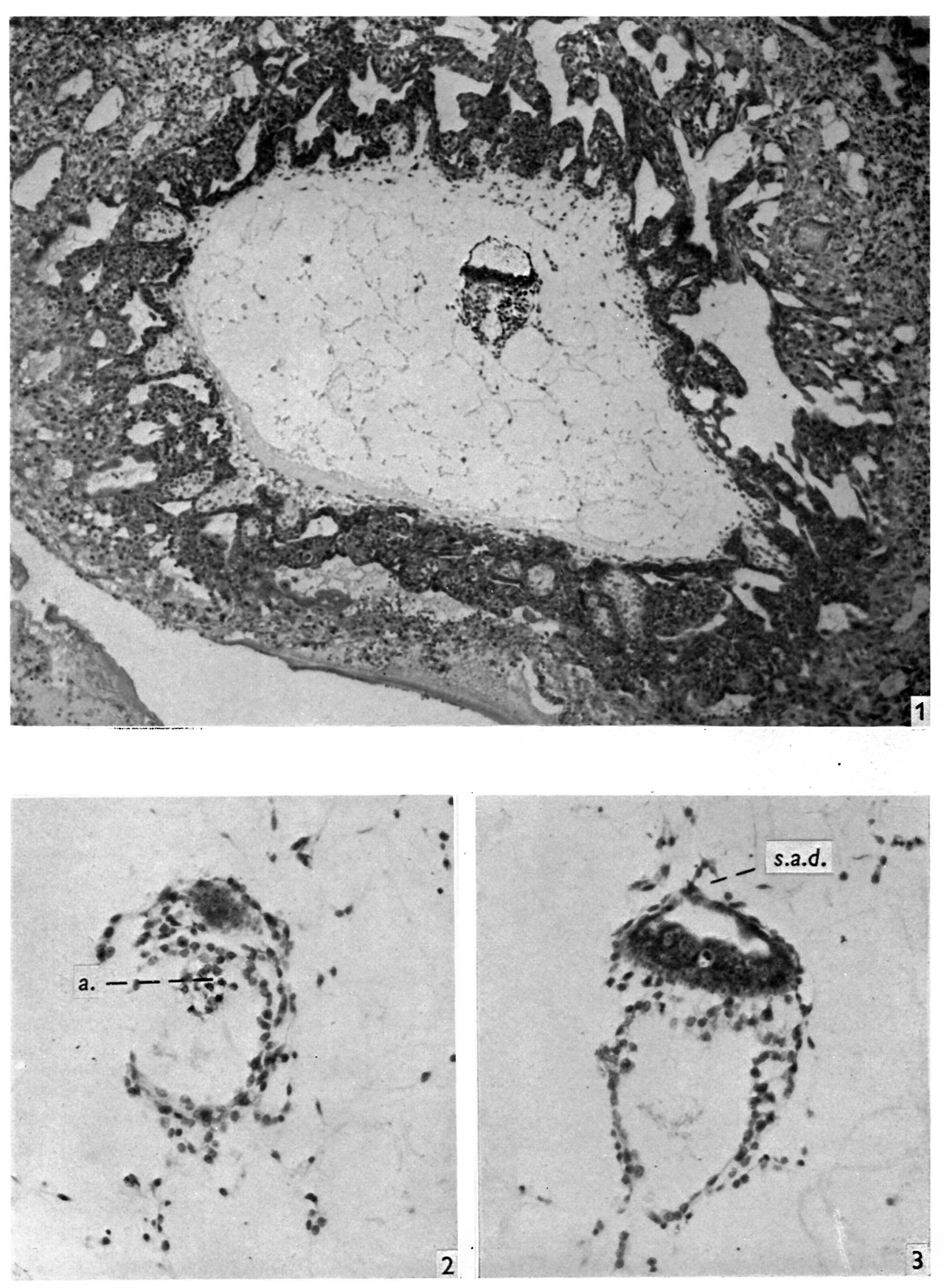 | 1,280 × 1,746 (405 KB) | Z8600021 (talk | contribs) | |
| 21:35, 6 August 2017 | 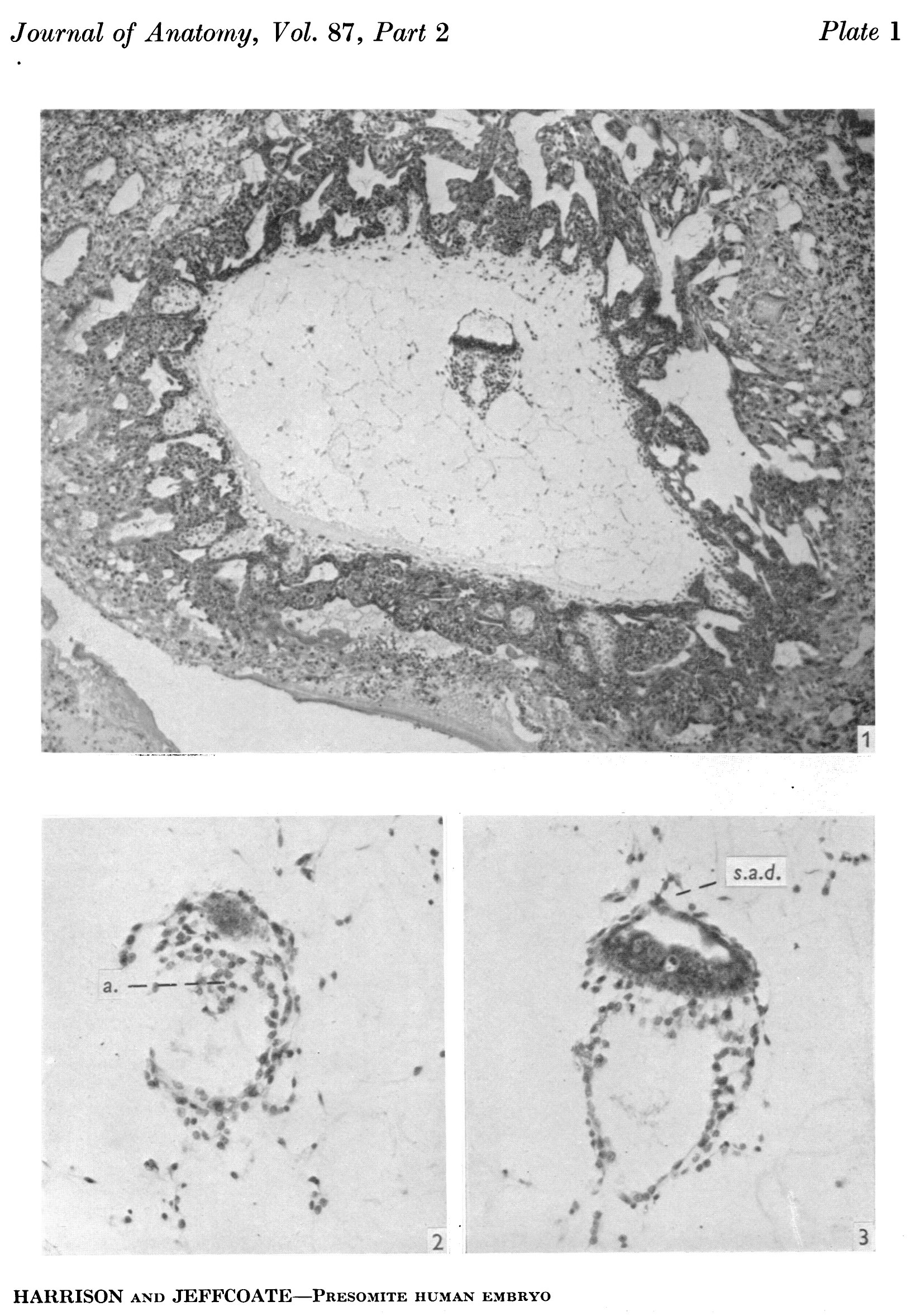 | 1,647 × 2,355 (747 KB) | Z8600021 (talk | contribs) |
You cannot overwrite this file.
File usage
The following page uses this file: