File:Fawcett1923 fig01.jpg: Difference between revisions
(==Fig 1== (after Bolk) Bolk illustrated his communication by four coloured figures which represent different ages in sequence. In fig. 1 one sees from below upwards, cartilage centres in the cervical vertebrae, separate on the two sides but connect...) |
mNo edit summary |
||
| (One intermediate revision by the same user not shown) | |||
| Line 9: | Line 9: | ||
{{Ref-Fawcett1923}} | {{Ref-Fawcett1923}} | ||
{{Footer}} | |||
[[Category:Skull]] | [[Category:Skull]] | ||
Latest revision as of 16:21, 30 January 2018
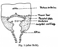
Fig 1
(after Bolk)
Bolk illustrated his communication by four coloured figures which represent different ages in sequence. In fig. 1 one sees from below upwards, cartilage centres in the cervical vertebrae, separate on the two sides but connected together dorsally by a spinal membrane; above the vertebrae on each side one sees cartilage in a somewhat broad sheet forming-what appears to be the side of a foramen magnum, and connected together about their middle by a narrow bar of cartilage from which a small process ascends. Below this bar is an uncoloured area, Bolk’s spino-occipital membrane, which is continuous with the spinal membrane below and contains two small nodules of cartilage. Above the transverse bar above mentioned is a transversely placed separate mass of cartilage.
Reference
Fawcett E. Some observations on the roof of the primordial human cranium. (1923) J Anat. 57(3): 245-250. PMID 17103974
Cite this page: Hill, M.A. (2024, June 24) Embryology Fawcett1923 fig01.jpg. Retrieved from https://embryology.med.unsw.edu.au/embryology/index.php/File:Fawcett1923_fig01.jpg
- © Dr Mark Hill 2024, UNSW Embryology ISBN: 978 0 7334 2609 4 - UNSW CRICOS Provider Code No. 00098G
File history
Yi efo/eka'e gwa ebo wo le nyangagi wuncin ye kamina wunga tinya nan
| Gwalagizhi | Nyangagi | Dimensions | User | Comment | |
|---|---|---|---|---|---|
| current | 18:38, 19 June 2017 |  | 749 × 585 (62 KB) | Z8600021 (talk | contribs) | |
| 18:38, 19 June 2017 |  | 767 × 650 (67 KB) | Z8600021 (talk | contribs) | ==Fig 1== (after Bolk) Bolk illustrated his communication by four coloured figures which represent different ages in sequence. In fig. 1 one sees from below upwards, cartilage centres in the cervical vertebrae, separate on the two sides but connect... |
You cannot overwrite this file.
File usage
The following page uses this file: