File:Fawcett1911 fig04.jpg: Difference between revisions
From Embryology
(Z8600021 uploaded a new version of File:Fawcett1911 fig04.jpg) |
mNo edit summary |
||
| Line 1: | Line 1: | ||
==Fig. 4. Coronal section Minot 37 mm embryo== | |||
I.N., supraorbltal nerve; P.M., palatine process of maxilla; T.R., tooth ridge. | |||
Latest revision as of 22:39, 22 May 2017
Fig. 4. Coronal section Minot 37 mm embryo
I.N., supraorbltal nerve; P.M., palatine process of maxilla; T.R., tooth ridge.
| Historic Disclaimer - information about historic embryology pages |
|---|
 |
Reference
Fawcett E. The development of the human maxilla, vomer, and paraseptal cartilages. (1911) J Anat. Physiol. 45(4): 378-405.
Cite this page: Hill, M.A. (2024, June 25) Embryology Fawcett1911 fig04.jpg. Retrieved from https://embryology.med.unsw.edu.au/embryology/index.php/File:Fawcett1911_fig04.jpg
- © Dr Mark Hill 2024, UNSW Embryology ISBN: 978 0 7334 2609 4 - UNSW CRICOS Provider Code No. 00098G
File history
Yi efo/eka'e gwa ebo wo le nyangagi wuncin ye kamina wunga tinya nan
| Gwalagizhi | Nyangagi | Dimensions | User | Comment | |
|---|---|---|---|---|---|
| current | 22:39, 22 May 2017 | 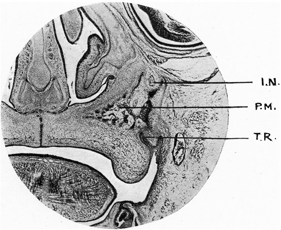 | 900 × 738 (182 KB) | Z8600021 (talk | contribs) | |
| 22:38, 22 May 2017 |  | 1,111 × 930 (201 KB) | Z8600021 (talk | contribs) |
You cannot overwrite this file.
File usage
The following page uses this file: