File:Patten1938 text-fig01.jpg: Difference between revisions
No edit summary |
(Z8600021 uploaded a new version of File:Patten1938 text-fig01.jpg) |
||
| (One intermediate revision by the same user not shown) | |||
| Line 1: | Line 1: | ||
==Text-Fig.1. Semischematic drawings of the interior of the heart to show the steps in its partitioning== | |||
(From Embryology, Patten, B. M., courtesy P. Blakiston’s Son and Co.) | |||
A. The cardiac septa are represented at the stage reached in human embryos early in the 5th week of development. Note especially the primary relations of interatrial septum primum. Based on original reconstructions of the heart of a 3.7 mm pig embryo, and on Tandler’s reconstructions of corresponding stages of the human heart. | |||
B. The cardiac septa as they appear in human embryos of the 6th week. Note the restriction of interatrial foramen primum by the growth of interatrial septum primum. Based on original reconstructions of the heart of a 6 mm pig embryo, on Born’s reconstructions of the rabbit heart, and Tandler’s reconstructions of corresponding stages of the human heart. | |||
===Reference=== | |||
{{Ref-Patten1938}} | |||
{{Footer}} | |||
Latest revision as of 16:49, 27 February 2017
Text-Fig.1. Semischematic drawings of the interior of the heart to show the steps in its partitioning
(From Embryology, Patten, B. M., courtesy P. Blakiston’s Son and Co.)
A. The cardiac septa are represented at the stage reached in human embryos early in the 5th week of development. Note especially the primary relations of interatrial septum primum. Based on original reconstructions of the heart of a 3.7 mm pig embryo, and on Tandler’s reconstructions of corresponding stages of the human heart.
B. The cardiac septa as they appear in human embryos of the 6th week. Note the restriction of interatrial foramen primum by the growth of interatrial septum primum. Based on original reconstructions of the heart of a 6 mm pig embryo, on Born’s reconstructions of the rabbit heart, and Tandler’s reconstructions of corresponding stages of the human heart.
Reference
Patten BM. Developmental defects at the foramen ovale. (1938) Am J Pathol. 14(2):135-162. PMID 19970381
Cite this page: Hill, M.A. (2026, October 16) Embryology Patten1938 text-fig01.jpg. Retrieved from https://embryology.med.unsw.edu.au/embryology/index.php/File:Patten1938_text-fig01.jpg
- © Dr Mark Hill 2026, UNSW Embryology ISBN: 978 0 7334 2609 4 - UNSW CRICOS Provider Code No. 00098G
File history
Yi efo/eka'e gwa ebo wo le nyangagi wuncin ye kamina wunga tinya nan
| Gwalagizhi | Nyangagi | Dimensions | User | Comment | |
|---|---|---|---|---|---|
| current | 16:49, 27 February 2017 | 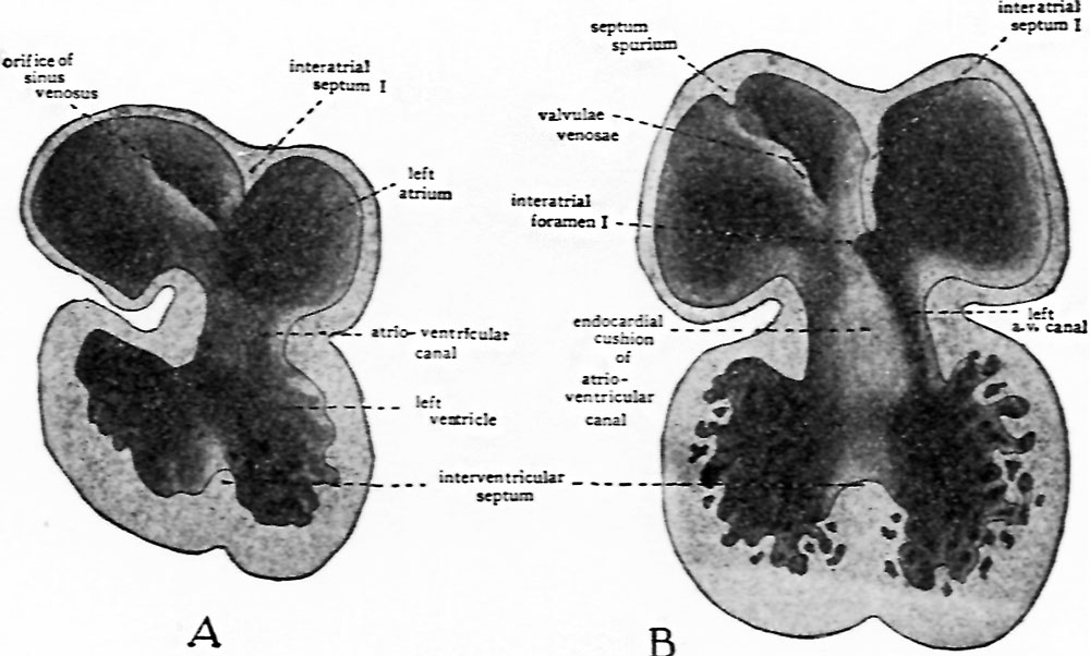 | 1,000 × 602 (99 KB) | Z8600021 (talk | contribs) | |
| 16:48, 27 February 2017 |  | 1,284 × 1,355 (338 KB) | Z8600021 (talk | contribs) |
You cannot overwrite this file.
File usage
The following page uses this file:


