File:Streeter1957 plate03.jpg: Difference between revisions
mNo edit summary |
|||
| (One intermediate revision by the same user not shown) | |||
| Line 3: | Line 3: | ||
Rudiments of the ducts are present in the youngest age group. Figure 24 reveals a condensed field of mesoderm beneath the epithelial bud. The latter invades the mesodermal bed and by repeated branchings forms the epithelial component of the gland. | Rudiments of the ducts are present in the youngest age group. Figure 24 reveals a condensed field of mesoderm beneath the epithelial bud. The latter invades the mesodermal bed and by repeated branchings forms the epithelial component of the gland. | ||
Fig. 22. No. 6524, xviii, 23-3-2 x200 | Fig. 22. [[:Category:Carnegie Embryo 6524|'''No. 6524''']], xviii, 23-3-2 x200 | ||
Fig. 23. No. 7707, xviii, 21-1-3, x200 | Fig. 23. [[:Category:Carnegie Embryo 7707|'''No. 7707''']], xviii, 21-1-3, x200 | ||
Fig. 24. No. 6824, xix, 12-1-4 x200 | Fig. 24. [[:Category:Carnegie Embryo 6824|'''No. 6824''']], xix, 12-1-4 x200 | ||
Fig. 25. No. | Fig. 25. [[:Category:Carnegie Embryo 1134B|'''No. 1134B''']], xx, 6-2-2 x150 | ||
{{Streeter1957 figures}} | {{Streeter1957 figures}} | ||
[[Category:Week 8]][[Category:Gland]] | [[Category:Week 8]][[Category:Gland]] | ||
[[Category:Carnegie Embryo 6524]][:Category:Carnegie Embryo 7707]][[Category:Carnegie Embryo 6824]][[Category:Carnegie Embryo 1134B]] | |||
Latest revision as of 18:09, 6 November 2016
Plate 3. Photographs of the Submandibular gland in embryos of horizons xvii, xix, and xx
Rudiments of the ducts are present in the youngest age group. Figure 24 reveals a condensed field of mesoderm beneath the epithelial bud. The latter invades the mesodermal bed and by repeated branchings forms the epithelial component of the gland.
Fig. 22. No. 6524, xviii, 23-3-2 x200
Fig. 23. No. 7707, xviii, 21-1-3, x200
Fig. 24. No. 6824, xix, 12-1-4 x200
Fig. 25. No. 1134B, xx, 6-2-2 x150
| Historic Disclaimer - information about historic embryology pages |
|---|
 |
- Links: 1 Graph Embryos 11-23 | 4 Eye and optic nerve 19-23 | Plate 1 - Cornea | Plate 2 - Hypophysis
Reference
Streeter GL. Developmental Horizons In Human Embryos Description Or Age Groups XIX, XX, XXI, XXII, And XXIII, Being The Fifth Issue Of A Survey Of The Carnegie Collection. (1957) Carnegie Instn. Wash. Publ. 611, Contrib. Embryol., 36: 167-196.
Cite this page: Hill, M.A. (2026, October 4) Embryology Streeter1957 plate03.jpg. Retrieved from https://embryology.med.unsw.edu.au/embryology/index.php/File:Streeter1957_plate03.jpg
- © Dr Mark Hill 2026, UNSW Embryology ISBN: 978 0 7334 2609 4 - UNSW CRICOS Provider Code No. 00098G[:Category:Carnegie Embryo 7707]]
File history
Yi efo/eka'e gwa ebo wo le nyangagi wuncin ye kamina wunga tinya nan
| Gwalagizhi | Nyangagi | Dimensions | User | Comment | |
|---|---|---|---|---|---|
| current | 17:50, 30 October 2016 | 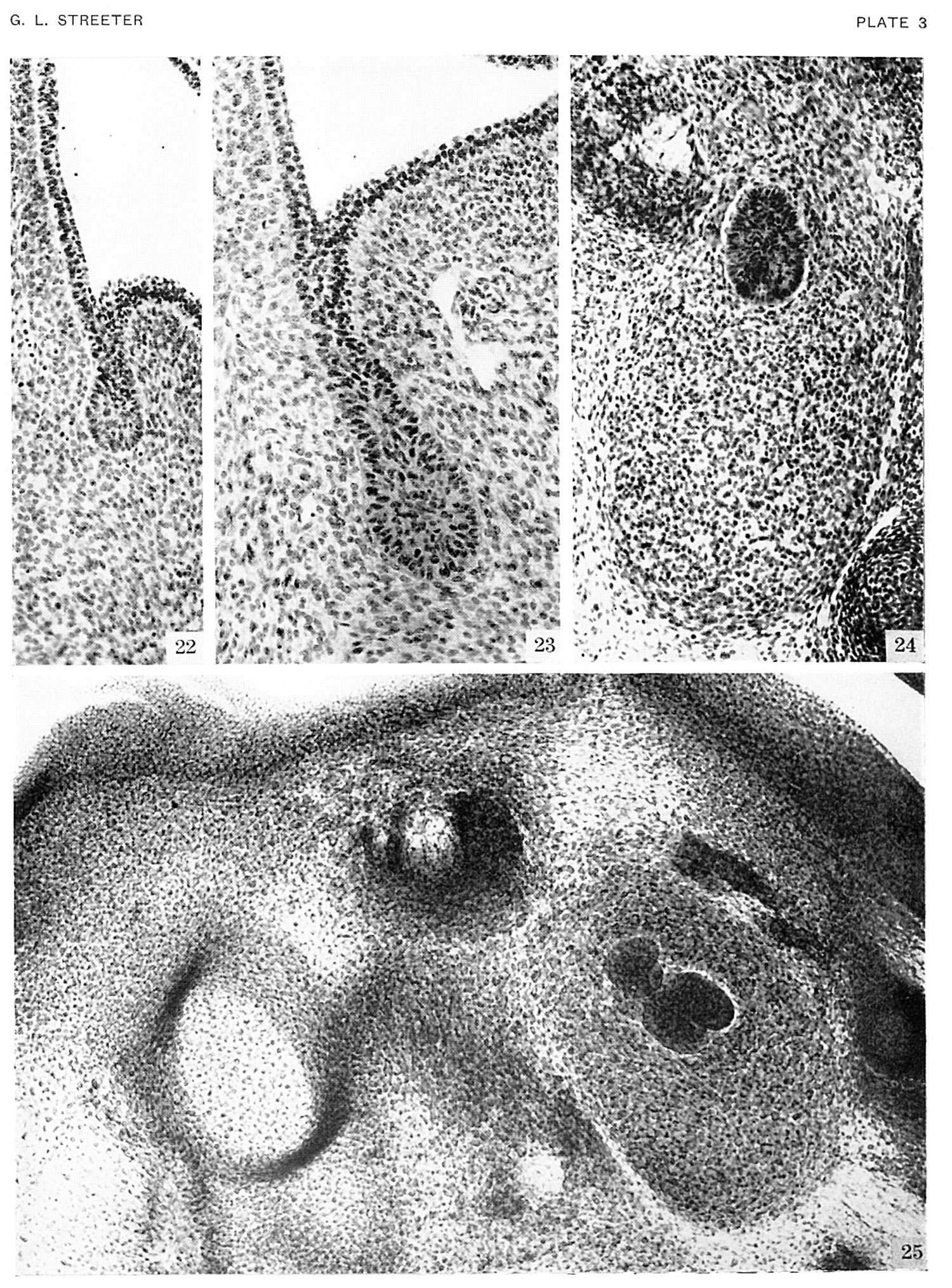 | 1,500 × 2,031 (892 KB) | Z8600021 (talk | contribs) |
You cannot overwrite this file.
File usage
The following page uses this file:


