File:Florian1930-text-fig08-09.jpg: Difference between revisions
No edit summary |
mNo edit summary |
||
| (4 intermediate revisions by the same user not shown) | |||
| Line 1: | Line 1: | ||
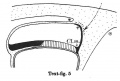
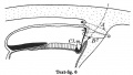
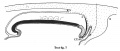
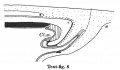
==Text-figs. 8-9. Schemes of the chief stages in the development of the human embryo== | |||
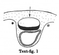
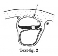
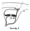
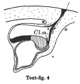
Ectoderm black; entoderm lined horizontally, primitive streak vertically, head process and chorda plate obliquely. Mesoderm dotted. The axis of the connecting stalk is marked by an arrow. The limits of the connecting stalk are marked by '''*''' and '''ʘ''', the limits of the umbilical stalk by '''+''' and '''ʘ'''). The direction of the extension of the amniotic cavity towards the connecting stalk is marked by arrows. The parts of the allantois and of the axis of the connecting stalk situated out of the median plane are finely dotted. A. Connecting stalk; B. umbilical stalk; Cl.m. cloacal membrane. | |||
<gallery> | |||
Florian1930-text-fig01.jpg|Text-fig 1 | |||
Florian1930-text-fig02.jpg|Text-fig 2 | |||
Florian1930-text-fig03.jpg|Text-fig 3 | |||
Florian1930-text-fig04.jpg|Text-fig 4 | |||
Florian1930-text-fig05.jpg|Text-fig 5 | |||
Florian1930-text-fig06.jpg|Text-fig 6 | |||
Florian1930-text-fig07.jpg|Text-fig 7 | |||
Florian1930-text-fig08.jpg|Text-fig 8 | |||
Florian1930-text-fig09.jpg|Text-fig 9 | |||
</gallery> | |||
{{Florian1930 figures}} | |||
Latest revision as of 19:36, 23 September 2015
Text-figs. 8-9. Schemes of the chief stages in the development of the human embryo
Ectoderm black; entoderm lined horizontally, primitive streak vertically, head process and chorda plate obliquely. Mesoderm dotted. The axis of the connecting stalk is marked by an arrow. The limits of the connecting stalk are marked by * and ʘ, the limits of the umbilical stalk by + and ʘ). The direction of the extension of the amniotic cavity towards the connecting stalk is marked by arrows. The parts of the allantois and of the axis of the connecting stalk situated out of the median plane are finely dotted. A. Connecting stalk; B. umbilical stalk; Cl.m. cloacal membrane.
Text-fig 1
Text-fig 2
Text-fig 3
Text-fig 4
Text-fig 5
Text-fig 6
Text-fig 7
Text-fig 8
Text-fig 9


| Historic Disclaimer - information about historic embryology pages |
|---|
 |
- Links: Text-fig 1 | Text-fig 2 | Text-fig 3 | Text-fig 4 | Text-fig 5 | Text-fig 6 | Text-fig 7 | Text-fig 1-7 | Text-fig 8 | Text-fig 9 | Text-fig 8-9 | Florian 1930 | Historic Embryology Papers
Reference
Florian J. The formation of the connecting stalk and the extension of the amniotic cavity towards the tissue of the connecting stalk in young human embryos. (1930) J. Anat., 64: 454-476.
Cite this page: Hill, M.A. (2024, June 26) Embryology Florian1930-text-fig08-09.jpg. Retrieved from https://embryology.med.unsw.edu.au/embryology/index.php/File:Florian1930-text-fig08-09.jpg
- © Dr Mark Hill 2024, UNSW Embryology ISBN: 978 0 7334 2609 4 - UNSW CRICOS Provider Code No. 00098G
File history
Yi efo/eka'e gwa ebo wo le nyangagi wuncin ye kamina wunga tinya nan
| Gwalagizhi | Nyangagi | Dimensions | User | Comment | |
|---|---|---|---|---|---|
| current | 19:11, 23 September 2015 |  | 1,000 × 1,347 (165 KB) | Z8600021 (talk | contribs) | |
| 19:11, 23 September 2015 | 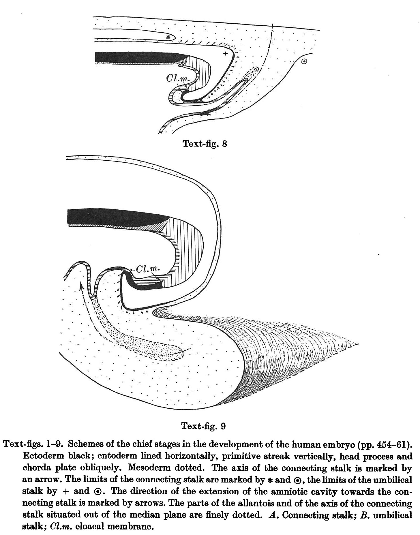 | 1,456 × 1,855 (357 KB) | Z8600021 (talk | contribs) |
You cannot overwrite this file.
File usage
The following page uses this file: