File:Wyndham1943 plate02.jpg: Difference between revisions
mNo edit summary |
m (→Plate 2) |
||
| Line 3: | Line 3: | ||
'''Fig. 5.''' Transverse section through caudal region of a 20 mm. embryo H 304. x 20. b, plica. inguinalis; g, pubic anlage. | '''Fig. 5.''' Transverse section through caudal region of a 20 mm. embryo H 304. x 20. b, plica. inguinalis; g, pubic anlage. | ||
'''Fig. 6.''' The same section as above. X120. Showing the detailed relations of the cells of the | '''Fig. 6.''' The same section as above. X120. Showing the detailed relations of the cells of the definitive {{gubernaculum}} to the belly wall. | ||
'''Fig. 7.''' Transverse section of a 42 mm. embryo H 42. x 11. a, testis; b, gubernaculum. | '''Fig. 7.''' Transverse section of a 42 mm. embryo H 42. x 11. a, testis; b, {{gubernaculum}}. | ||
'''Fig. 8.''' Transverse section of a 42 mm. embryo H 42. x 100. c, cremaster muscle; d, processus vaginalis. | '''Fig. 8.''' Transverse section of a 42 mm. embryo H 42. x 100. c, cremaster muscle; d, processus vaginalis. | ||
Latest revision as of 15:41, 7 June 2019
Plate 2
Fig. 5. Transverse section through caudal region of a 20 mm. embryo H 304. x 20. b, plica. inguinalis; g, pubic anlage.
Fig. 6. The same section as above. X120. Showing the detailed relations of the cells of the definitive gubernaculum to the belly wall.
Fig. 7. Transverse section of a 42 mm. embryo H 42. x 11. a, testis; b, gubernaculum.
Fig. 8. Transverse section of a 42 mm. embryo H 42. x 100. c, cremaster muscle; d, processus vaginalis.
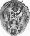
Fig. 5. Transverse section through caudal region of a 20 mm embryo H 304
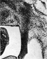
Fig. 6. Gubernaculum detail 20 mm embryo H 304
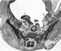
Fig. 7. Transverse section of a 42 mm embryo H 42
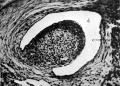
Fig. 8. Transverse section of a 42 mm embryo H 42


| Historic Disclaimer - information about historic embryology pages |
|---|
 |
- Links: Plate 1 | Fig 1 | Fig 2 | Fig 3 | Fig 4 | Plate 2 | Fig 5 | Fig 6 | Fig 7 | Fig 8 | Plate 3 | Fig 9 | Fig 10 | Fig 11
Reference
Wyndham NR. A morphological study of testicular descent. (1943) J Anat., 77(2):179-188.3. PMID 17104926
Cite this page: Hill, M.A. (2024, June 5) Embryology Wyndham1943 plate02.jpg. Retrieved from https://embryology.med.unsw.edu.au/embryology/index.php/File:Wyndham1943_plate02.jpg
- © Dr Mark Hill 2024, UNSW Embryology ISBN: 978 0 7334 2609 4 - UNSW CRICOS Provider Code No. 00098G
File history
Click on a date/time to view the file as it appeared at that time.
| Date/Time | Thumbnail | Dimensions | User | Comment | |
|---|---|---|---|---|---|
| current | 19:17, 4 September 2015 | 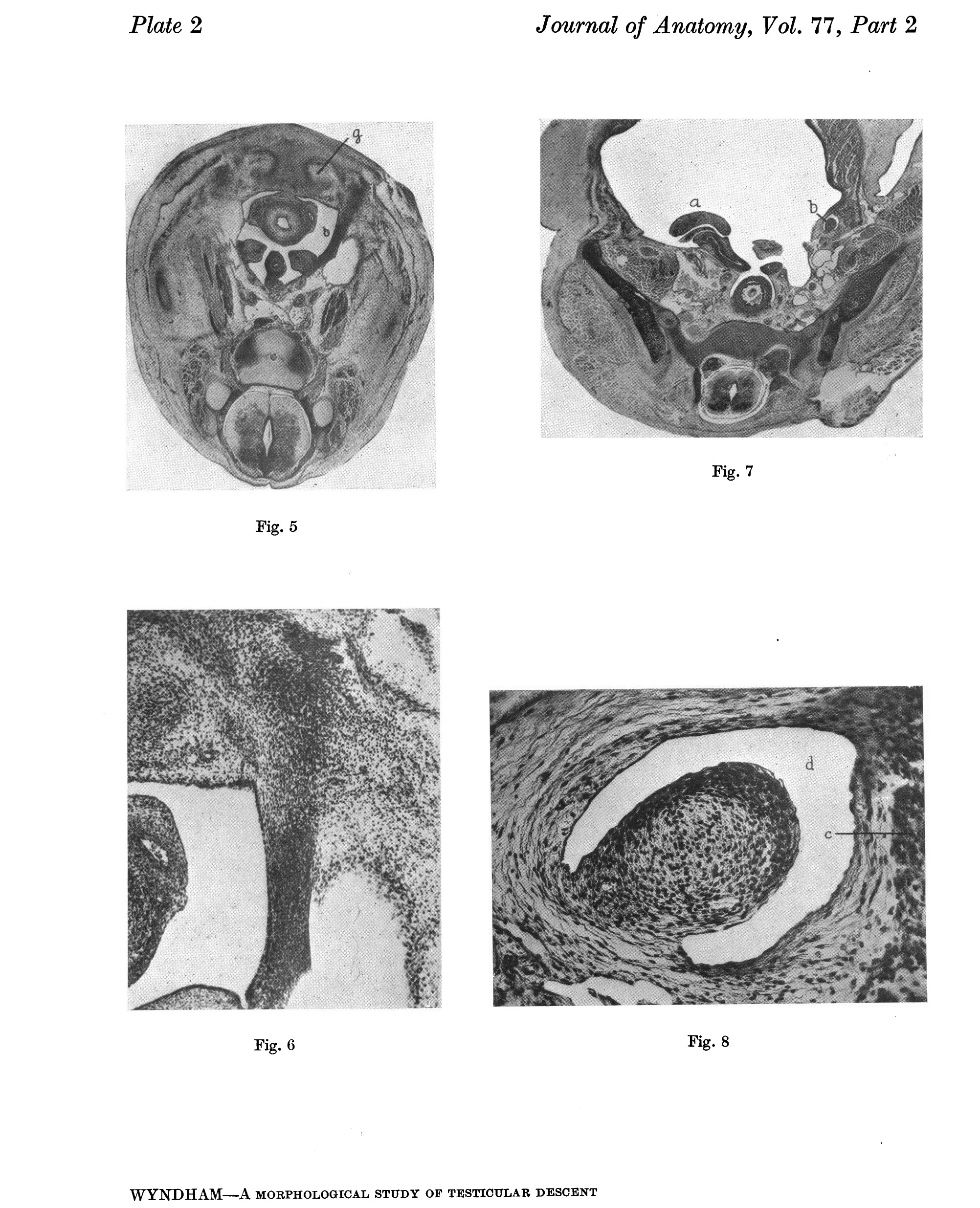 | 2,004 × 2,471 (990 KB) | Z8600021 (talk | contribs) |
You cannot overwrite this file.
File usage
The following page uses this file: