File:Dynamic Localization of Two Membrane Proteins for Fertilization.jpg: Difference between revisions
No edit summary |
No edit summary |
||
| Line 2: | Line 2: | ||
=Dynamic Localization of Two Membrane Proteins Required for Fertilization= | =Dynamic Localization of Two Membrane Proteins Required for Fertilization= | ||
(A-D) Localization of SPE-9 in spermatids (A and B) and spermatozoa (C and D). SPE-9 localizes to the plasma membrane of spermatids. After spermiogenesis, SPE-9 is enriched at the plasma membrane of the pseudopod. Photographs reproduced from Zannoni et al. (2004), and used with permission. (E) High pressure freezing transmission electron microscopy of spermatozoa (s) in the spermatheca. Membranous organelles (MO) and pseudopods (ps) are indicated. During spermiogenesis (not shown) the MOs fuse with the plasma membrane and maintain a persistant fusion pore, flanked by an electron-dense collar. Electron micrograph courtesy of Kent McDonald. Bar, 1 μm.<ref><pubmed>18050412</pubmed></ref> | (A-D) Localization of SPE-9 in spermatids (A and B) and spermatozoa (C and D). SPE-9 localizes to the plasma membrane of spermatids. After spermiogenesis, SPE-9 is enriched at the plasma membrane of the pseudopod. Photographs reproduced from Zannoni et al. (2004), and used with permission. (E) High pressure freezing transmission electron microscopy of spermatozoa (s) in the spermatheca. Membranous organelles (MO) and pseudopods (ps) are indicated. During spermiogenesis (not shown) the MOs fuse with the plasma membrane and maintain a persistant fusion pore, flanked by an electron-dense collar. Electron micrograph courtesy of Kent McDonald. Bar, 1 μm.<ref><pubmed>18050412</pubmed></ref> | ||
Latest revision as of 00:28, 15 August 2015
Dynamic Localization of Two Membrane Proteins Required for Fertilization
(A-D) Localization of SPE-9 in spermatids (A and B) and spermatozoa (C and D). SPE-9 localizes to the plasma membrane of spermatids. After spermiogenesis, SPE-9 is enriched at the plasma membrane of the pseudopod. Photographs reproduced from Zannoni et al. (2004), and used with permission. (E) High pressure freezing transmission electron microscopy of spermatozoa (s) in the spermatheca. Membranous organelles (MO) and pseudopods (ps) are indicated. During spermiogenesis (not shown) the MOs fuse with the plasma membrane and maintain a persistant fusion pore, flanked by an electron-dense collar. Electron micrograph courtesy of Kent McDonald. Bar, 1 μm.[1]
[original figure legend : Meioticfig3_s.jpg]
References
- ↑ <pubmed>18050412</pubmed>
Copyright
© 2005 David Greenstein. This is an open-access article distributed under the terms of the Creative Commons Attribution License, which permits unrestricted use, distribution, and reproduction in any medium, provided the original author and source are credited.
- Note - This image was originally uploaded as part of an undergraduate science student project and may contain inaccuracies in either description or acknowledgements. Students have been advised in writing concerning the reuse of content and may accidentally have misunderstood the original terms of use. If image reuse on this non-commercial educational site infringes your existing copyright, please contact the site editor for immediate removal.
File history
Yi efo/eka'e gwa ebo wo le nyangagi wuncin ye kamina wunga tinya nan
| Gwalagizhi | Nyangagi | Dimensions | User | Comment | |
|---|---|---|---|---|---|
| current | 23:15, 14 August 2015 | 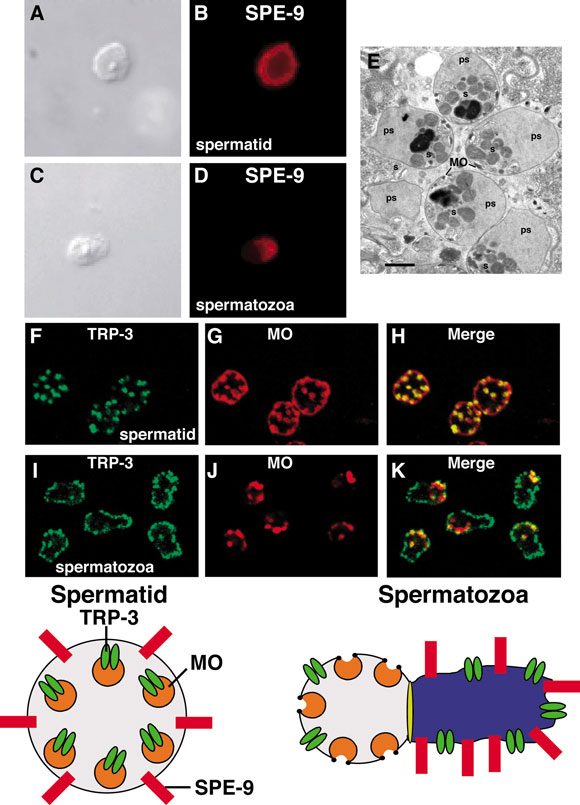 | 580 × 805 (88 KB) | Z3463890 (talk | contribs) | Meioticfig3_s.jpg PMID 18050412 |
You cannot overwrite this file.
File usage
The following page uses this file: